Fig. 5.
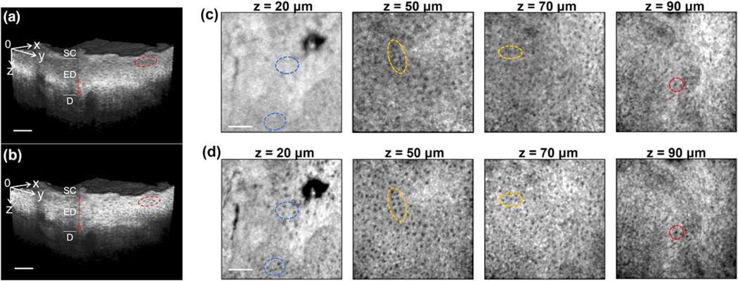
Human skin epidermis. Gaussian beam was focused at 90 μm depth. 80 μm x 1.5 μm NB started at z = 20 μm and ended at z = 100 μm. (a) Volumetric data of Gaussian beam imaging. The bright and narrow layer marked by the red double-arrow line is within DOF. The cells in the red circle are fuzzy. (b) 3D data captured by 80 μm N B. (c), (d) XY images at five depths demonstrate the resolution disparity between Gaussian beam and 80 μm NB. Cells highlighted in red ellipses have good visibility with both beams while the cells in yellow ellipses are clear in 80 μm NB imaging but noisy in Gaussian beam imaging. Cells in blue ellipses are visible with 80 μm NB but completely disappear with Gaussian beam imaging. Scale bar, 100 μm; XY = 0.5 mm × 0.5 mm; SC, stratum corneum; ED, epidermis; D, dermis.

