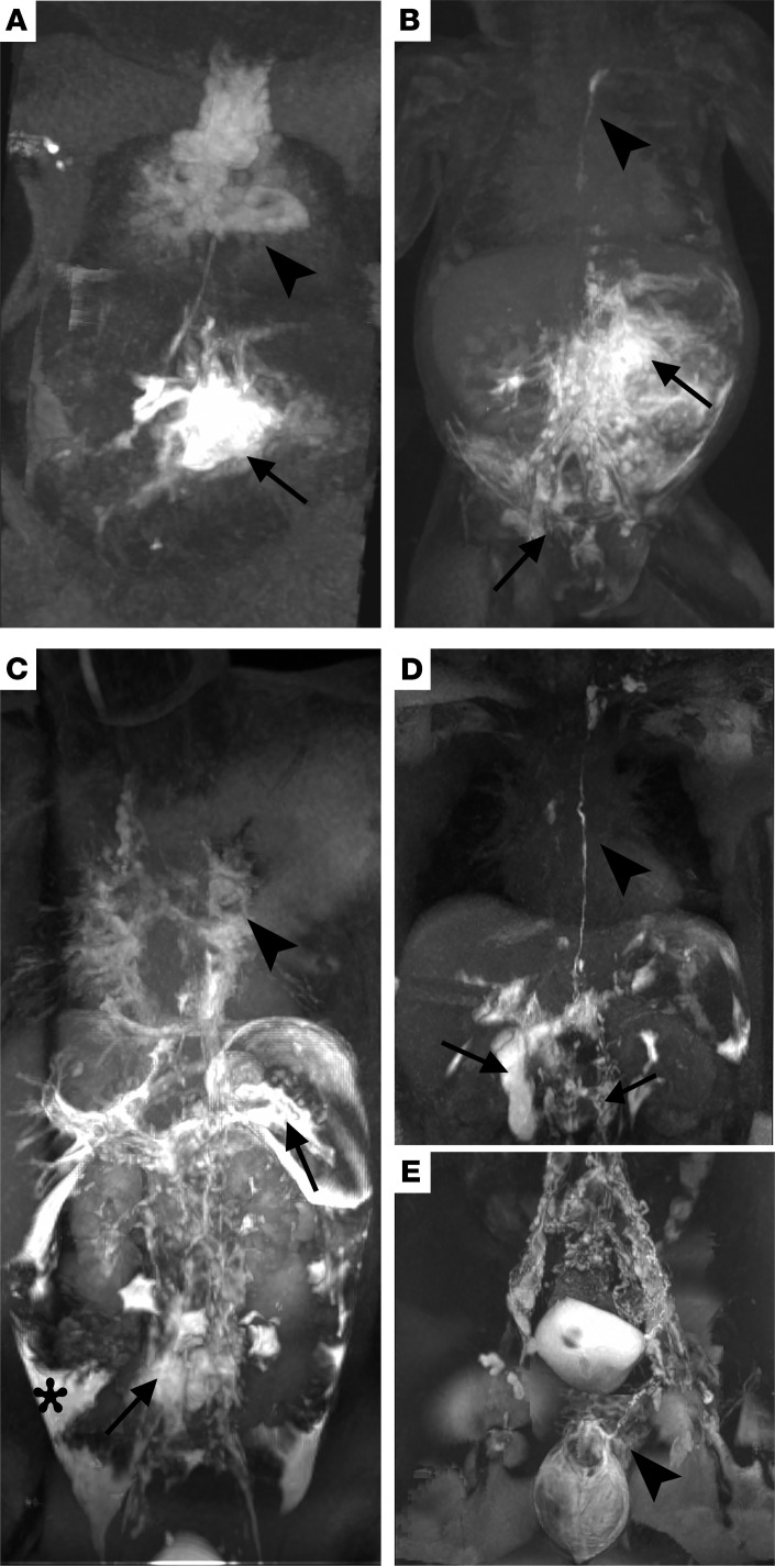
Figure 1. DCMRL of individuals 1, 2, and 4.
(A) Intrahepatic DCMRL of individual 1 illustrating retrograde mesenteric perfusion (arrow) and pulmonary lymphatic perfusion with dilated mediastinal lymphatics (arrowhead). (B) Intranodal DCMRL of individual 1 shows retrograde into the mesentery (arrow), right renal, and dermal lymphatics (arrow), with an i.p. leak and intact thoracic duct coursing to the left venous angle (arrowhead). (C) Intrahepatic DCMRL of individual 2 shows retrograde flow into the lumbar and iliac lymphatics (arrow), splenic lymphatics (arrow), and i.p. leak (*). There is also bilateral pulmonary and mediastinal lymphatic perfusion (arrowhead) without a thoracic duct. (D) Intrahepatic and (E) intranodal DCMRL of individual 4 show a normal-appearing thoracic duct (arrowhead) with retrograde lumbar perfusion (arrow) and intraduodenal leak (arrow). Intranodal DCMRL shows dilated iliac lymphatics with retrograde dermal perfusion (scrotal and penile).

