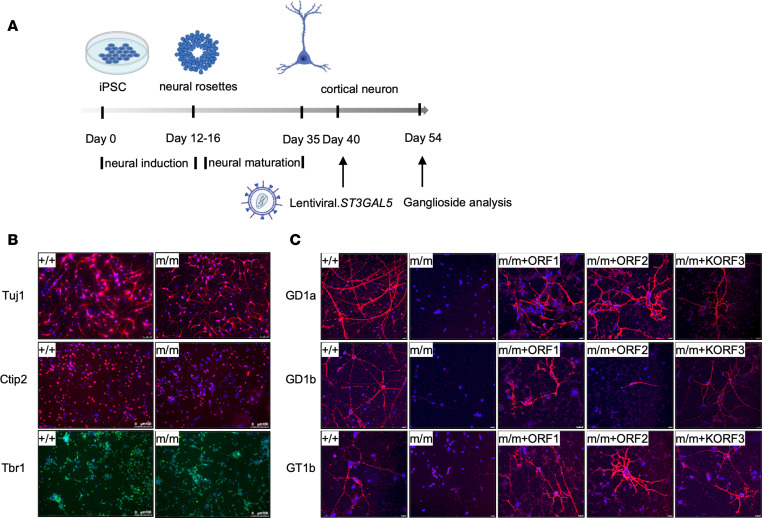
Figure 2. ST3GAL5 replacement restores gangliosides production in iPSC-derived cortical neurons.
(A) Workflow to examine restoration of ganglioside production in patients’ iPSC differentiated cortical neurons by lentiviral vectors expressing ST3GAL5 ORFs. (B) Representative images of neuronal markers in ST3GAL5+/+ and ST3GAL5mut/mut iPSC-differentiated cortical neurons. Neuron-specific class III β-tubulin (Tuj1) and COUP-TF-interacting protein 2 (Ctip2) are indicated by red; T-box brain transcription factor 1 (TBR1) is indicated by green, with nuclei counterstained in blue. (C) Representative images of major brain gangliosides in cortical neurons by lentiviral vectors expressing ST3GAL5 ORFs. GD1a, GD1b, and GT1b are indicated by red; nuclei are counterstained in blue. +/+, WT; m/m, ST3GAL5mut/mut. Scale bars: 100 µm (B), 10 µm (C).

