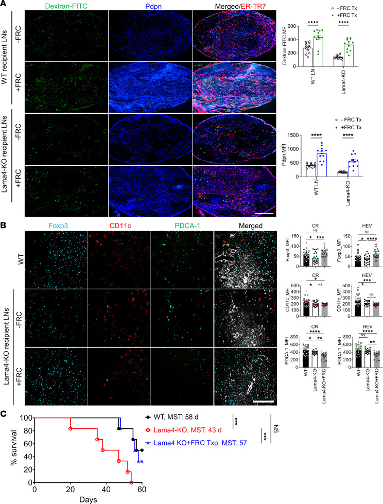
Figure 6. FRC transfer restores FRC-Lama4-KO lymph node impairments and allograft acceptance.
WT FRCs (1 × 10⁵) were injected i.v. into WT or FRC-Lama4-KO mice weekly for 4 weeks. One week after the fourth dose, conduit systems were visualized 90 minutes after injecting 40 kDa dextran-FITC. (A) Left: Whole-mount scanning fluorescence images of LN cryosections from WT and FRC-Lama4-KO mice with and without FRC transfer. Scale bar: 500 μm. Right: Quantification of dextran-FITC and Pdpn in LNs. (B) FoxP3+ Tregs, CD11c+ cDCs, and Pdca-1+ pDCs in LNs. Scale bar: 100 μm. Data in A and B are representative of 3 independent experiments; 3 mice/group, 5 LNs/mouse, 3 sections/LN, 3–5 fields/section. (C) One week after the fourth dose of FRC, Lama4-KO mice received cardiac transplants from BALB/c mice, 250 μg anti-CD40L mAb i.v. on day 0, and weekly WT FRCs after transplantation (1 × 10⁵ FRCs/dose/week); 6 recipients/group. Allograft survival was monitored for 8 weeks. WT and FRC-Lama4-KO recipients without FRC transfer were used as controls. Data presented as mean ± SEM. *P < 0.05; **P < 0.01; ****P < 0.001; ****P < 0.0001 by 1-way ANOVA with Tukey’s multiple-comparison test (A and B) or 2-tailed log-rank (Mantel-Cox) test (C).

