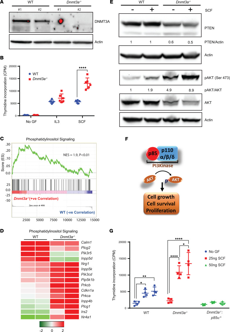
Figure 1. Loss of Dnmt3a in BMNC results in increased cell proliferation via the PI3K pathway.
(A) WT and Dnmt3afl/fl:Mx-Cre mice were administered poly I:C at 12–16 weeks of age, and BMNCs were subjected to Western blot analysis to detect the presence of DNMT3a protein. Two independent WT and Dnmt3a–/– mouse–derived BMNCs were utilized for these experiments. (B) BMNCs from WT or Dnmt3a–/– mice were cultured in media supplemented with SCF (50 ng/mL) or IL-3 (10 ng/mL) or no growth factor for 48 hours. Cell proliferation was evaluated by [3H] thymidine incorporation. Counts per minute (CPM) are shown. Three independent experiments, n = 6, mean ± SD, ****P < 0.00005. BM cells collected from WT or Dnmt3a–/– mice were subjected to RNA isolation and, subsequently, next-generation sequencing. (C and D) GSEA revealed an enrichment for genes in the PI3K signaling pathway (C), and upregulation of specific genes in the PI3K pathway are shown in the heatmap (D). (E) BMNCs from WT or Dnmt3a–/– mice were starved of growth factors and stimulated with SCF, followed by Western blot analysis. (F) Class I PI3K complex is composed of p85 regulatory subunit and p110α, p110β, and p110δ catalytic subunits. Activated PI3K signaling regulates AKT phosphorylation, which in turn promotes cell grow, cell survival, and proliferation. (G) WT, Dnmt3afl/fl:Mx-Cre, and Dnmt3afl/fl:p85αfl/fl:Mx-Cre mice were administered poly I:C at 12–16 weeks of age. BMNCs were collected from 3 different mice of each genotype and stimulated with SCF (25 ng/mL or 50 ng/mL) or in the absence of growth factors for 48 hours. Cell proliferation was evaluated by [3H] thymidine incorporation. Counts per minute (CPM) are shown. Experiment performed 3 times, and representative experiment shown with n = 4, mean ± SD, *P < 0.05, **P < 0.005, ****P < 0.00005. Two-way ANOVA, Tukey’s multiple-comparison test (B and C).

