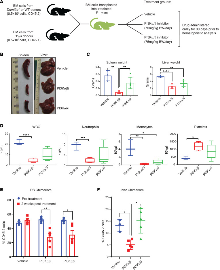
Figure 2. PI3K αβ inhibition modulates Dnmt3a loss–induced myeloid leukemia development.
(A) Donor cells from Dnmt3a–/– mice were mixed with BoyJ cells in 1:1 ratio (0.5 × 10⁶ versus 0.5 × 10⁶) for a competitive transplantation assay. Six weeks after transplantation, mice were treated with vehicle or the PI3K αβ inhibitor (Bay1082439; 7 mg/kg body weight) or PI3K αδ inhibitor (GDC-0941; 75 mg/kg body weight) for 30 days, and mice were analyzed. (B) Representative images of liver and spleen from vehicle- and drug-treated mice are shown. (C) Quantitative assessment of spleen and liver weights from the indicated groups. n = 3–5, mean ± SD, **P < 0.005, ****P < 0.00005. (D) PB counts from mice described in B and C before they were sacrificed. n = 3–5, *P < 0.05, **P < 0.005, ***P < 0.0005, ****P < 0.00005. The boxes shown with lower and upper quartiles separated by the median (horizontal line), and the whiskers extend to the minimum and maximum values. (E and F) Mice described in A were analyzed for PB chimerism 2 weeks after drug treatment and after 30 days after drug treatment for liver chimerism. Chimerism was assessed by staining the cells using an anti-CD45.2 antibody and flow cytometry. n = 3–5, mean ± SEM, *P < 0.05, **P < 0.005. One-way ANOVA in C, D, and F; 2-way ANOVA in E with Tukey’s multiple comparison test performed.

