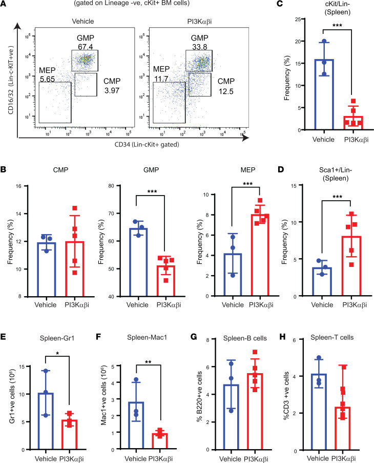
Figure 4. PI3K αβ inhibition decreases myeloid progenitors and improves megakaryocyte-erythrocyte progenitors in Dnmt3a–/– malignant mice.
(A and B) Flow cytometry analysis was performed on BM cells from mice transplanted with Dnmt3a–/– cells and treated with the PI3K αβ inhibitor. Representative dot plots and quantitative data showing the frequency of Lin–/c-KIT+ myeloid progenitors: CMPs, GMPs, and MEPs. n = 3–5, mean ± SEM, ***P = 0.0005. (C and D) Spleen cells collected from vehicle- or PI3Kαβ inhibitor–treated mice as in A were subjected to flow cytometry analysis to detect Lin–c-KIT+Sca-1+ cells. Quantitative data showing reduced c-KIT+ (Lin–) myeloid progenitor cells (C) and increased differentiated c-KIT– spleen cells (D) in PI3K αβ inhibitor–treated group compared with controls. n = 3–5, mean ± SEM, ***P = 0.0005. (E–H) Flow cytometry was performed on spleen cells from vehicle or PI3K αβ inhibitor–treated mice as in A. Quantitative data showing a reduction in myeloid cell burden in the spleen from drug treated Dnmt3a–/– mice compared with controls. n = 3–5, mean ± SEM, *P = 0.05, **P = 0.005, unpaired t test (2-tailed) performed (B–H).

