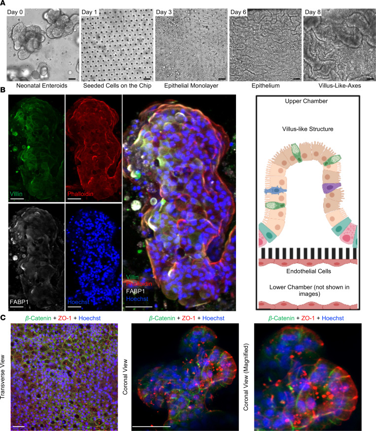
Figure 1. Development of the Neonatal-Intestine-on-a-Chip microfluidic model.
(A) Growth progression of Neonatal-Intestine-on-a-Chip is shown by brightfield microscopy, beginning with neonatal enteroids on day 0, seeding of stem cells within a microfluidic device on day 1, development of a confluent monolayer by day 3, invaginations of epithelium through day 6, and advancement to villus-like-axes on day 8. Scale bars: 50 μm. Images representative of more than 20 independent experiments. (B) Left: Representative immunofluorescence images of a deconvoluted coronal cross section of a villus-like formation within the Neonatal-Intestine-on-a-Chip is shown separated and merged (merged) with villin (green, microvilli brush border), phalloidin (red, actin), FABP1 (white, enterocytes), and Hoechst (blue, nuclei). Scale bars: 50 μm. Right: Illustration demonstrating coronal view seen in immunofluorescence images (created with BioRender.com). (C) Representative immunofluorescence images of Neonatal-Intestine-on-a-Chip epithelium stained for β-catenin (green, basolateral component of adherens junctions), ZO-1 (red, apical tight junctions), and with Hoechst (blue). Scale bars: 50 μm.

