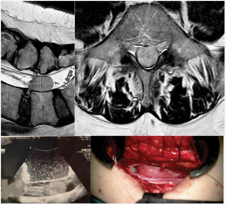
Figure 2.
(A) Preoperative T2-MR image of an intradural space-occupying lesion at level L5, showing (B) homogenous contrast enhancement on T1-MR imaging. This lesion was a lumbal spinal solitary fibrous tumor, also termed hemangiopericytoma, WHO II, which reflects one of the possible differential diagnoses when a spinal meningioma is suspected in preoperative MR imaging. (C) The intraoperative ultrasound image confirmed the correct level and extent of surgical access before dural opening. (D) In contrast to most spinal meningiomas, this tumor showed a more reddish coloration during surgery, which is indicative of higher vascularization.

