
Fig. 1. TSC tauopathy is a neurofibrillary tauopathy with prominent tau acetylation.
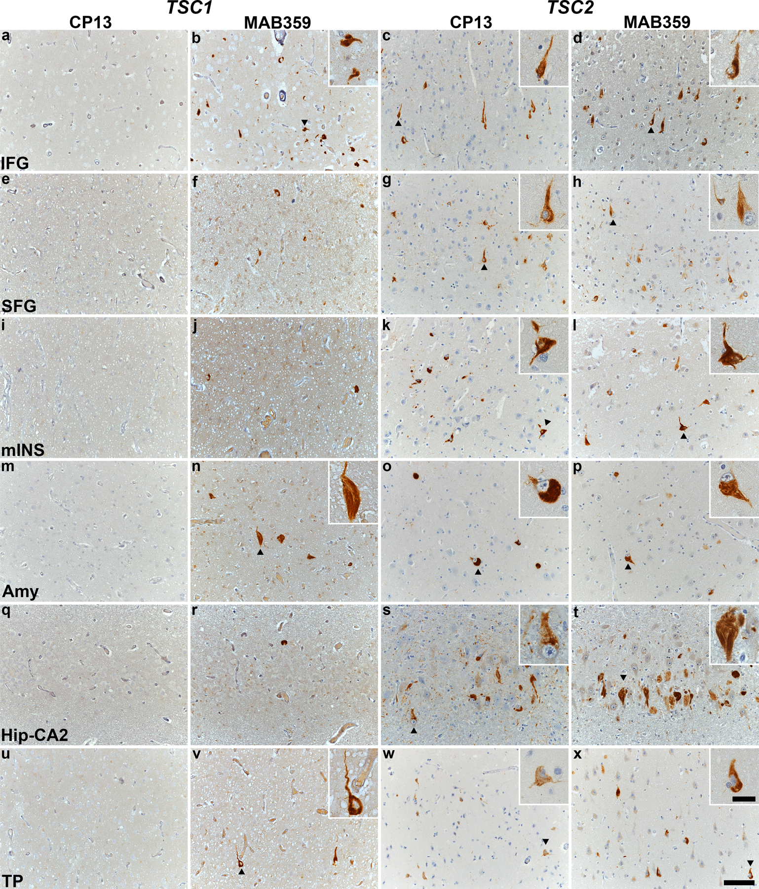
Immunohistochemical staining with CP13 and MAB359 antibodies in TSC subjects carrying pathogenic variants in TSC1 (TS 6, a-b, e-f, i-j, m-n, q-r, u-v) and TSC2 (TS 10, c-d, g-h, k-l, o-p, s-t, w-x). Representative images of immunostaining for CP13 and MAB359 in inferior frontal gyrus (IFG, a-d), superior frontal gyrus (SFG, e-h), middle insula (mINS, i-l), amygdala (Amy, m-p), hippocampal CA2 (Hip-CA2, q-t) and temporal pole (TP, u-x) are shown, highlighting neurofibrillary tangles (arrowheads) in the insets. All sections were counterstained with hematoxylin. Scale bars in panel x are 100 µm and 25 µm (inset) and apply to all images.