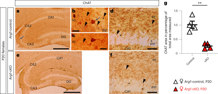
Fig. 6. The Arg1-cKO hippocampi of 2- to 3-month-old female mice receive reduced cholinergic innervation.
a–f, Microphotographs of sagittal sections of Arg1-control (a–d) and Arg1-cKO (e,f) P20 female hippocampi. ChAT immunoreactivity was revealed by using DAB as a chromogen. Triangular or ovoid immunoreactive ChAT interneurons were observed in the CA3 field of the Arg1-control hippocampus (a,b, arrows). Cholinergic axons show the characteristic varicosities (b–d,f, arrowheads) of the boutons of en passant synapses. At the pyramidal cell layer (pyr), immunoreactive fibers delineate the pyramidal neuronal somata (so; d,f, asterisk). g, Quantitative analysis demonstrates that the female Arg1-cKO hippocampus receives less cholinergic innervation than the Arg1-control hippocampus; P = 0.0016. Each triangle corresponds to one animal (n = 4). Scale bars, 500 µm (a,e), 50 µm (d,f) and 20 µm (b,c). Data are shown as mean ± s.e.m. Statistically significant differences were determined by an unpaired two-sided t-test; **P ≤ 0.01.

