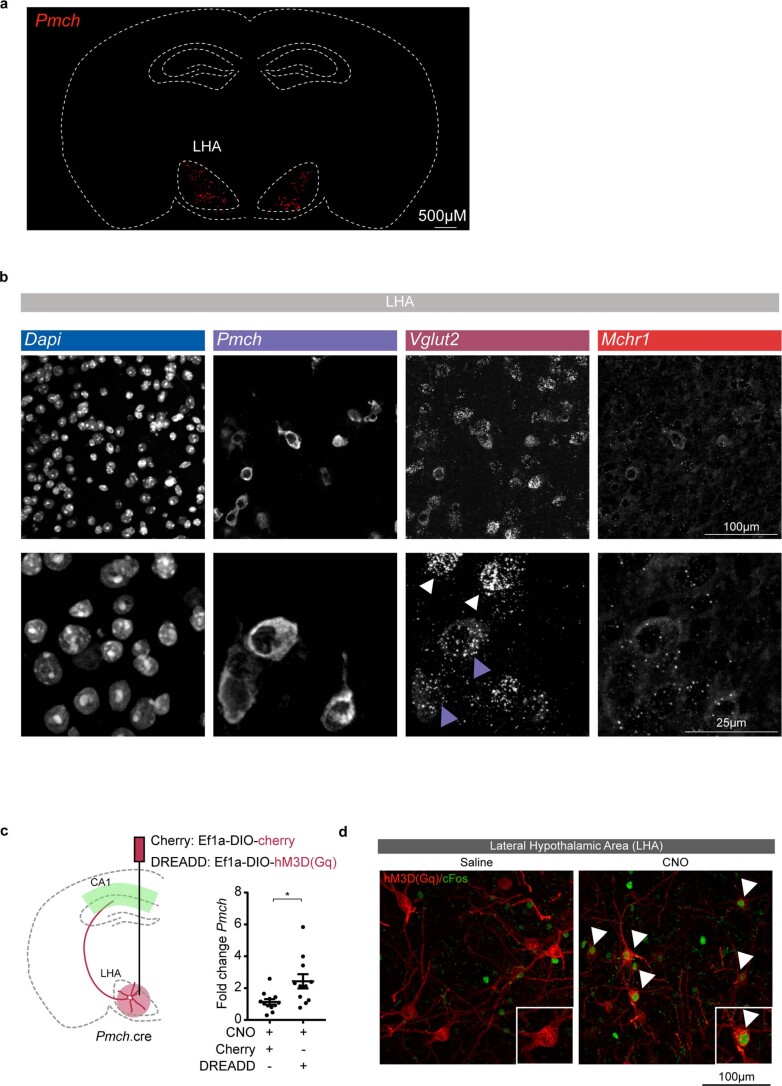
Extended Data Fig. 5. Pmch is expressed in LHA cell-bodies.
a,b, RNAscope on 10μm Wt mouse brain coronal section showing (a) Pmch expression in whole section and (b) Pmch, Vglut2 (Slc17a6) and Mchr1 expression in the lateral hypothalamic area (LHA) (top row) and highlighted in crops (bottom row). White arrowheads indicate Vglut2 positive cells that do not express Pmch. Purple arrowheads indicate Vglut2- and Pmch-positive cells. c, RT-qPCR on hippocampal extracts from Pmch.cre animals expressing Cherry or hM3Dq(Gq) (DREADD-Cherry) in MCH-neurons in the LHA. CNO was i.p. injected at the beginning of the light-phase for 4 hours (3 mg/kg). Number of mice: Cherry n = 12, DREADD n = 11 (p = 0.0115). Two-tailed unpaired t-test. Individual data points shown with bars representing mean ± SEM. (*p < 0.05). d, Representative images from Pmch.cre animal expressing hM3Dq(Gq) in MCH-neurons in the LHA. Saline or CNO were i.p. injected at the beginning of the light-phase for 4 hours (3 mg/kg), to show that CNO injection induces cFos expression in hM3Dq(Gq)-expressing neurons. Arrowheads indicate active MCH-neurons that express hM3Dq(Gq) (red) and cFos (green). These animals belong to a different cohort from the animals used in Extended Data Fig. 5c. Number of mice: Saline n = 1, CNO n = 1.

