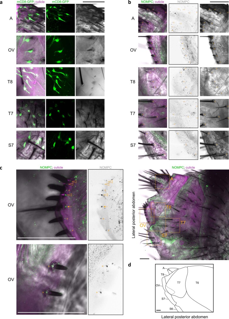
Extended Data Fig. 3. Mono-innervation and anti-NOMPC labeling of terminalia bristles.
a, Representative images of posterior abdomen from one of four pan-neuronal elav-GAL4>mCD8–GFP females. Left and middle: mCD8-GFP, pan-neuronal expression demonstrating the innervation of each bristle by the distal process of a single bipolar neuron (green). The innervation patterns associated with the long sensillum and short sensilla of the ovipositor (hypogynium) could not be deciphered. Left: autofluorescence, abdominal cuticle (magenta). Left and right: bright-field (grayscale). Here and in b-d, A, analia; OV, ovipositor valves; T8, 8th abdominal tergite; T7, 7th abdominal tergite; S7, 7th abdominal sternite; scale bar, 50 μm. b, Top five rows: representative images of posterior abdomen from one of nine wild-type female flies stained with anti-NOMPC (green at left, grayscale at middle). Left: autofluorescence, abdominal cuticle (magenta). Left and right: bright-field (grayscale). Arrowheads, foci of anti-NOMPC labeling observed at the base of all bristles34. Bottom: lower resolution image of the posterior abdomen, lateral aspect, containing the regions displayed above (orange boxes). c, Representative images of the ovipositor valves from two of nine wild-type flies stained with anti-NOMPC (green at left, grayscale at right). Left, autofluorescence, abdominal cuticle (magenta); bright-field (grayscale). Foci of anti-NOMPC labeling at the base of the three hypogynial short sensilla, the singular long sensillum, and the hypogynial teeth are indicated by triangles, the arrow, and the arrowheads, respectively. d, Diagram of female posterior abdomen, lateral aspect.

