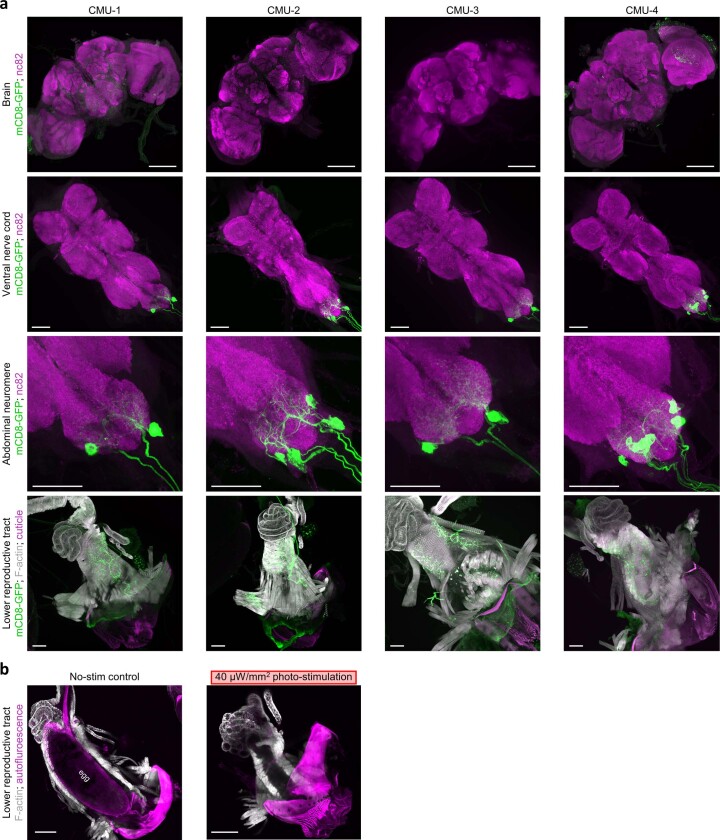
Extended Data Fig. 10. Expression pattern of CMU-splitGAL4 lines.
a, Representative images of the brain (top row), ventral nerve cord (second, third rows), and lower reproductive tract (bottom row) from CMU-splitGAL4>mCD8-GFP females, stained with anti-GFP (membrane of CMU neurons, green). Top three rows: tissue also stained with nc82 (synaptic neuropil, magenta). Bottom row: tissue also stained with phalloidin (muscle f-actin, gray); autofluorescence, abdominal cuticle (magenta). Representative brain images (top row) from 12, 2, 2, 6 flies. Representative ventral nerve cord images (second, third rows) from 8, 22, 6, 12 flies. Representative lower reproductive tract images (bottom row) from 8, 11, 2, 10 flies. Images in third row are higher resolution regions of images in second row. For quantitation of expression patterns in the ventral nerve cord and lower reproductive tract, see Supplementary Table 4. Here and in b, scale bar, 50 μm. b, Representative images of the lower reproductive tract from gravid CMU-4>CsChrimson females that were flash frozen in liquid nitrogen without (left, one of two flies) or with (right, one of four flies) concurrent photo-stimulation (655 nm light at 40 µW/mm2). Prior to experiment, gravid females were maintained in the dark in an environment that ensured egg retention in the uterus, as in Fig. 8d (Methods).

