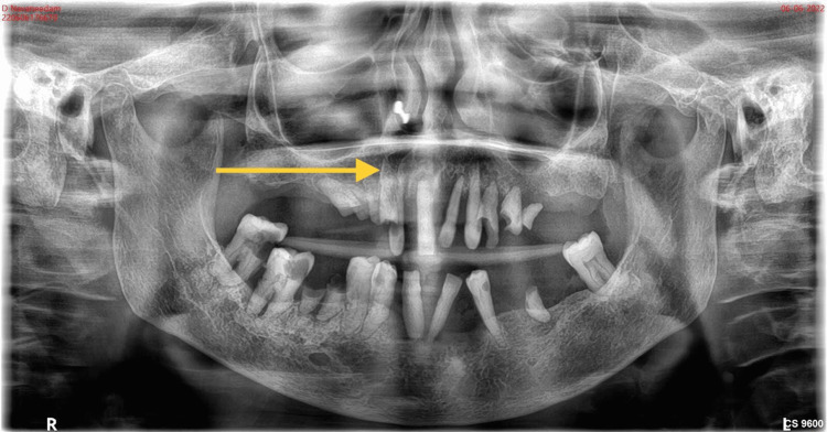
Figure 2. Pre-operative radiograph.
Represents the orthopantomogram showing the generalized horizontal bone loss in the upper arch and root stumps 13, 14, 15, 23, 24, 25, 36, and 46. Dental caries involving pulp in 47, 48, and 44 approximating pulp in 12 and 34. The arrow mark shows the generalized horizontal bone loss in the maxilla.

