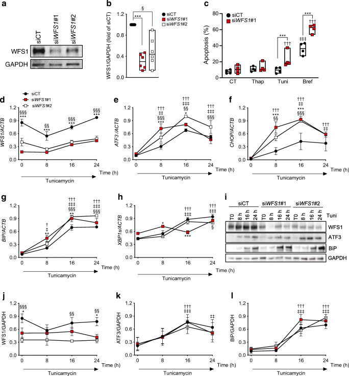
Fig. 3.
WFS1 silencing sensitises human beta cells to ER stress. WFS1 was silenced or not (siCT) in EndoC-βH1 cells using two siRNAs (siWFS1#1 and #2). (a) Representative western blot and (b) densitometric quantification of WFS1 knockdown 72 h after transfection. GAPDH was used as a control for protein loading. (c) Apoptosis assessed by Hoechst 33342/propidium iodide staining in control and WFS1-silenced EndoC-βH1 cells exposed or not for 24 h to thapsigargin, tunicamycin or brefeldin. Data points represent independent experiments. Extremities of floating bars are maximal and minimal values; horizontal line shows median. (d–l) Time course of tunicamycin exposure in EndoC-βH1 cells silenced for WFS1 for 48 h. WFS1, ATF3, CHOP, BIP and XBP1s mRNA expression was measured by real-time PCR (d–h) and normalised to reference gene ACTB. WFS1, ATF3 and BiP expression was examined by western blot and normalised to the reference protein GAPDH (i–l). Results are means ± SEM of n=3 (real-time PCR) or n=4 (western blots) independent experiments, and are expressed as fold of the highest value in each experiment. *p<0.05, **p<0.01, ***p<0.001 siWFS1#1 vs siCT, §p<0.05, §§p<0.01, §§§p<0.001 siWFS1#2 vs siCT; †p<0.05, ††p<0.01, †††p<0.001 treated vs CT or vs time 0 h in WFS1-deficient cells; ‡p<0.05, ‡‡p<0.01, ‡‡‡p<0.001 treated vs CT or vs time 0 h in control cells. Data were analysed by one-way or two-way ANOVA (as suitable), followed by Sidak’s or Dunnett’s test for multiple comparisons. Bref, brefeldin; CT, control untreated; Thap, thapsigargin; Tuni, tunicamycin

