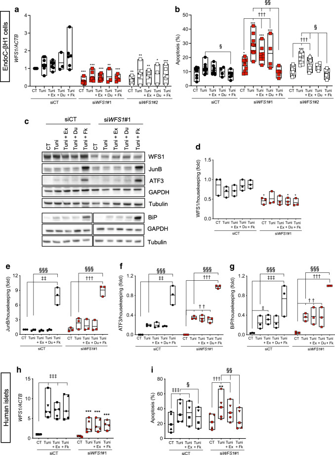
Fig. 4.
Exenatide protects human beta cells from ER stress-induced apoptosis. WFS1 was silenced in EndoC-βH1 cells (a–g) or dispersed human islets (h–i) using siRNAs (siWFS1#1 and #2). At 24 h after transfection, the cells were pretreated or not for 2 h or 24 h, respectively, with exenatide (50 nmol/l), dulaglutide (50 mmol/l) or forskolin (20 μmol/l), and then exposed or not (CT) for 24 (a, b), 16 (c–g) or 48 h (h, i), respectively, to tunicamycin alone or in combination with exenatide, forskolin or dulaglutide. (a, h) WFS1 mRNA expression by real-time PCR. (b, i) Apoptosis evaluated by Hoechst 33342/propidium iodide staining. (c–g) Western blot data. WFS1, JunB, ATF3 and BiP protein expression was normalised to the geometric mean of the reference proteins tubulin and GAPDH, and expressed as fold of the highest value in each experiment. Data points represent independent experiments. Extremities of the floating bars are maximal and minimal values; horizontal line shows median. *p<0.05, **p<0.01, ***p<0.001 siWFS1#1 or siWFS#2 vs the same treatment in siCT; ††p<0.01, †††p<0.001 treated vs CT in WFS1-silenced cells; ‡p<0.05, ‡‡p<0.01, ‡‡‡p<0.001 treated vs CT in control cells; §p<0.05, §§p<0.05, §§§p<0.001 Tuni+Ex or Tuni+Fk vs Tuni, by one-way ANOVA followed by Sidak’s or Tukey’s test for multiple comparisons. CT, control (vehicle); Du, dulaglutide; Ex, exenatide; Fk, forskolin; Tuni, tunicamycin

