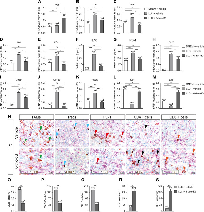
Fig. 7. Telomere dysfunction induced by 6-thio-dG treatment reduces expression of lung inflammation and tumor immunosupression markers.
Lung tissue mRNA expression levels of Ifng (anti-tumor immunity) (A), Tnf and Il1b (Th1 inflammation) (B, C) and Il10 and PD-1 (tumor immunosupression) (D, E) normalized to 18S expression, and IL10 (F) and PD-1 (G) protein levels in lung homogenates from LLC-challenged mice treated with 6-thio-dG vs. controls. Lung tissue mRNA expression levels of Ccl2 (macrophage chemotaxis) (H), Cd68 (tumor associated macrophages (TAMs)) (I), Cd163 (M2 TAMs) (J), Foxp3 (regulatory T cells (Tregs)) (K), Cd4 (CD4+ helper T cells) (L) and Cd8 (CD8+ cytotoxic T cells) (M) normalized to 18S expression in LLC-challenged mice treated with 6-thio-dG vs. controls. Representative lung immunostainings for PanCK (purple) with CD68 (brown; green arrowheads indicate CD68+ cells), FOXP3 (brown; blue arrowheads indicate FOXP3+ cells), PD-1 (brown; red arrowheads indicate PD-1+ cells), CD4 (brown; black arrowheads indicate CD4+ cells), and CD8 (brown; orange arrowheads indicate CD8+ cells) (N), and quantification of CD68 positive area (O), and number of FOXP3 (P), PD-1 (Q), CD4 (R) and CD8 (S) positive cells/mm2 in LLC-challenged mice treated with 6-thio-dG vs. controls. Data are expressed as mean ± SEM (the number of mice is indicated in each case). *p < 0.05; **p < 0.01; ***p < 0.001 (Dunn–Sidak test for multiple comparisons and Mann–Whitney or unpaired t tests to compare 2 independent groups).

