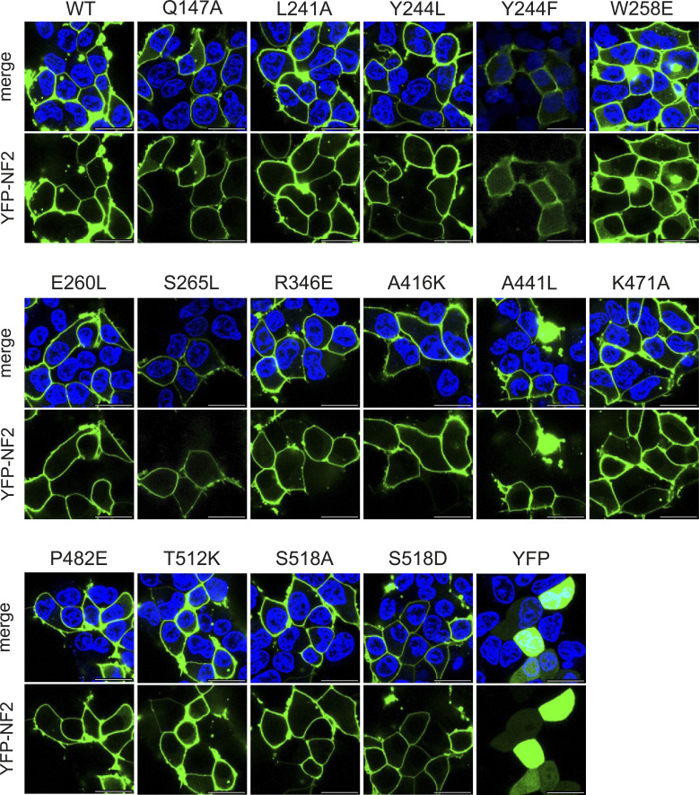
Figure S4. Confocal fluorescence microscopy images of wild-type and all tested NF2 variants.
(Related to Fig 5): as in Fig 5A. Confocal fluorescence microscopy images of WT and the full set of NF2 variants. HEK293T cells were transiently transfected with N-terminal YFP-tagged NF2 (green) and cellular localization of NF2 proteins 24 h after transfection was investigated by confocal fluorescence microscopy. Nuclei were stained with Hoechst (blue). Top, merge of Hoechst and YFP, bottom, YFP signal from NF2 expression. All variants were located at the membrane. Scale bar = 20 μM.

