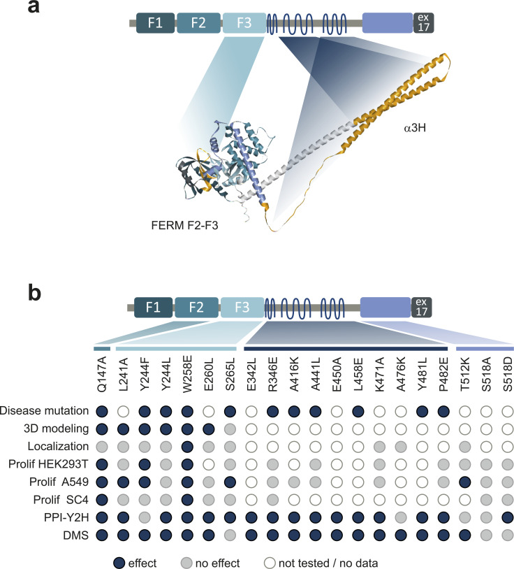
Figure S5. Overview of the functional assessment of NF2 variants affecting NF2 conformation and function.
(Related to the discussion): (A) schematic and 3D model of NF2 summarizing of regions critical for NF2 conformation. FERM-F3 and the α2H/α3H helix regions harboring mutations critical for conformation-dependent interactions are colored in orange in the alpha fold 3D structure (AF P35240 F1). (B) Summary of the NF2 variant effects. Disease mutations: a blue dot indicates an annotated SUV at the amino acid position. 3D modelling: a blue dot indicates a change of 3D interactions for the amino acid substitution. Localization: blue indicates a changed subcellular distribution for the amino acid substitution. Prolif: a blue dot indicates altered proliferation upon NF2 variant expression of HEK293T, A459 or SC4 cells, respectively. PPI-Y2H: a blue dot indicates alter protein interaction patterns in Y2H spot assays with individually cloned NF2 variants. DMS: a blue dot indicates interaction perturbation of the amino acid substitution in deep scanning experiments. Grey, no effect in comparison to WT; White/no fill, not tested.

