
Fig. 2.
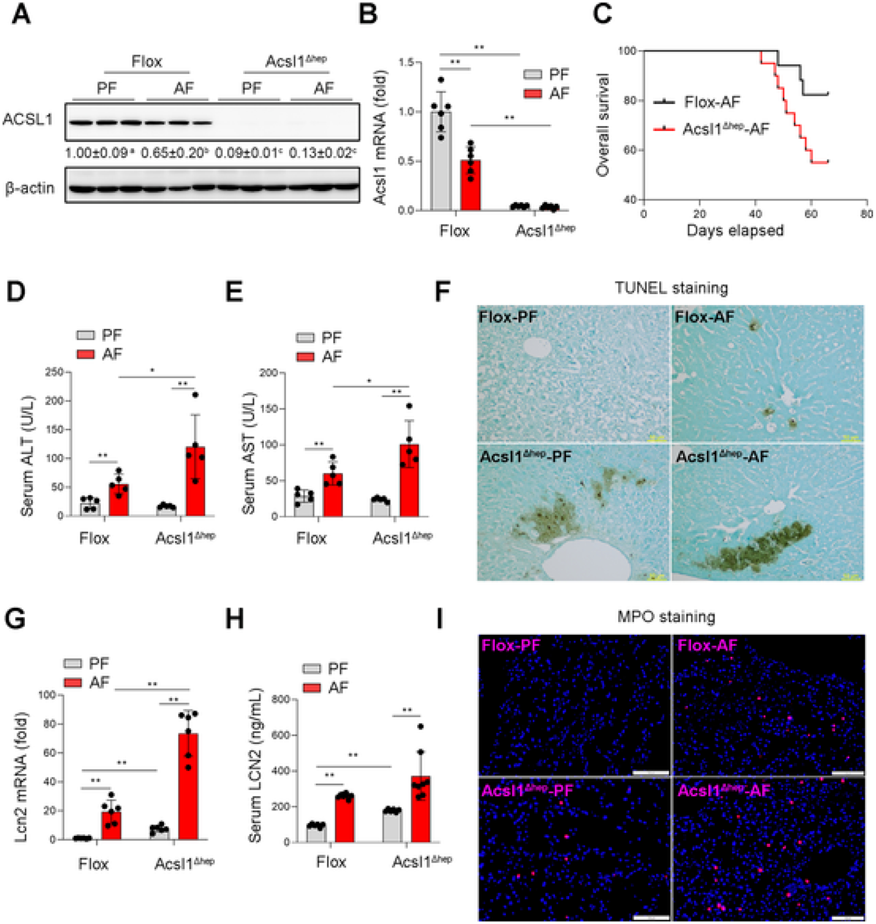
ACSL1 deficiency aggravates alcohol-induced liver injury. Wild-type Flox mice and hepatocyte-specific ACSL1 knockout mice (Acsl1 Δhep) were subjected to alcohol feeding for 8 weeks. (A) Western blot analysis of ACSL1 protein levels (n = 3/group). (B) qPCR assay of hepatic Acsl1 mRNA levels (n = 6/group). (C) Kaplan–Meier survival analysis determined the survival probability between the Flox mice and Acsl1Δhep mice with alcohol feeding for 8 weeks. (D–E) Serum levels of ALT (D) and AST (E) (n = 5/group). (F) TUNEL staining on the liver tissue sections. Scale bars = 50 μm. (G) qPCR assay of hepatic Lcn2 mRNA levels (n = 6/group). (H) Serum levels of LCN2 (n = 6–8/group). (I) IF staining of MPO in liver tissue sections (n = 6/group). Scale bars = 100 μm. Data are presented as mean ± SD. *P < 0.05, **P < 0.01, values with different superscripts are significantly different.