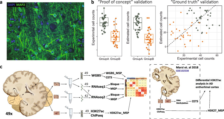
Fig. 1.
Study workflow. a IHC section in PFC showing the complexity of the human brain. DAPI staining (blue) was used to identify cell nuclei. MAP2 (green) is a neuronal marker expressed in neuronal bodies and processes. Neuronal processes present in the section may originate from the same cell as the nuclei, from nearby cells, or from projecting neurons whose bodies are located in an entirely different brain region. b The caveat of “proof of concept” validation for cell composition estimation methods. Simulated data, each point corresponds to one sample. The estimated counts (middle) show high performance based on “proof of concept” validation, recapitulating the group differences observed with experimental counts (left). However, direct comparison of the estimated and direct counts across samples (“Ground truth validation”) indicates poor correlation between the two, and failure of the estimates to correctly recapitulate the intra-group variability. c Study workflow. Performance of different estimation approaches was assessed through correlations among the estimates in same or nearby tissue samples from 49 individuals (left) and through re-analysis of H3K27ac ChIP-seq data with major differences in cell composition between the groups. Detailed description is provided under “Methods” section and Fig. 4a

