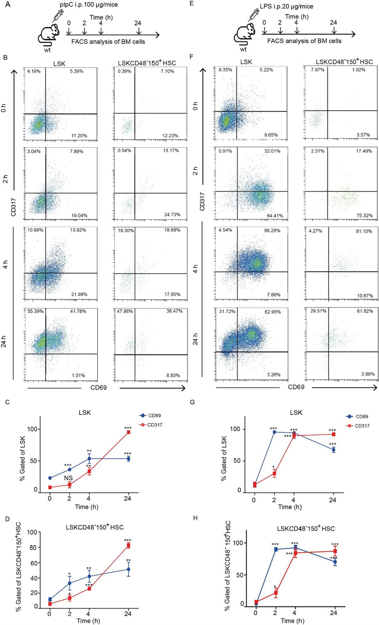
Fig. 1.
Immune activation of hematopoietic stem and progenitor cells is rapid. A Schematic representation of the experimental settings involving injection of pIpC at different time points (0, 2, 4, and 24 h). B Representative FACS plots showing the percentage of CD69+ and CD317+ cells in the LSK (left panels) and LSKCD48−150+ HSCs (right panels) populations from control and pIpC stimulated mice at different time points. C, D Quantification of CD69 and CD317 expression in the LSK and LSKCD48−150+ HSCs populations in the BM of control and pIpC stimulated mice. E Schematic representation of the experimental settings involving injection of LPS at different time points (0, 2, 4, and 24 h). F Representative FACS plots showing the percentage of CD69+ and CD317+ cells in the LSK (left panels) and LSKCD48−150+ HSCs (right panels) populations from control and LPS-stimulated mice at different time points. G, H Quantification of CD69 and CD317 expression in the LSK and LSKCD48−150+ HSCs populations in the BM of control and LPS-stimulated mice. *p < 0.05, **p < 0.01, ***p < 0.001

