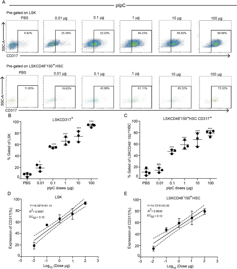
Fig. 2.
CD317 reveals a dose–response to pIpC above a minimal threshold. A Representative FACS plots showing the percentage of CD317+ cells in the LSK (upper panels) and LSKCD48−150+ HSCs (lower panels) populations from the BM of PBS-treated (control) and pIpC- stimulated mice under various pIpC doses (0.01–100 µg as positive control) at 24 h post-injection. B, C Quantification of the LSK CD317+ and LSKCD48−150+ HSCs CD317+ cell populations from the BM of PBS-treated (control) mice and mice treated with different doses of pIpC. D, E Linear regression model fitting the dose-dependent response with the expression of CD317 in LSK and LSKCD48−150+ HSCs populations. Solid lines represent the linear fit of data. Dotted lines represent 95% confidence intervals. Data are presented as average ± SD, a summary of three independent experiments, n ≥ 3 mice per group; *p < 0.05, **p < 0.01, ***p < 0.001

