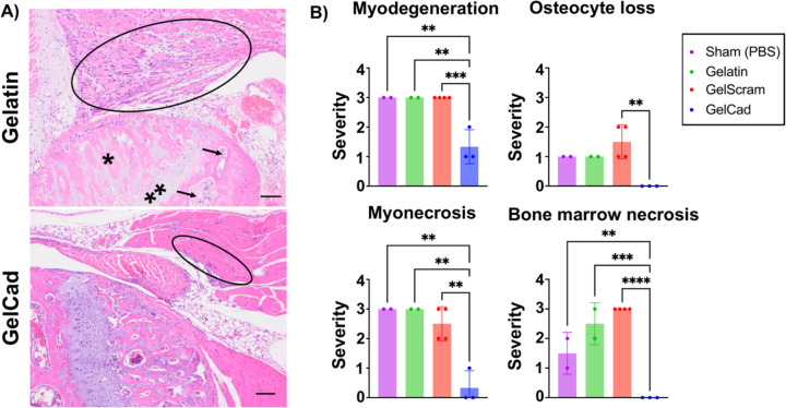
Figure 6: Histopathology of ligated hindlimbs receiving different hydrogel treatments.
A) Representative photomicrographs of H&E-stained images of the knee in ligated limbs of mice treated with gelatin versus GelCad hydrogels. In the gelatin image, marked myoregeneration with scattered myodegeneration and necrosis is encircled, and severe necrosis of the bone marrow (arrows), osteonecrosis (*), and chondronecrosis (**) are evident. In the GelCad image, only very mild myoregeneration is noted (encircled).
B) Quantification of pathology (0–3 scale). GelCad and GelScram samples were evaluated at day 14 post-ligation, and gelatin and sham samples were evaluated at day 7 post-ligation. Each data point represents the score from a single mouse. Data are presented as mean ± SD from N=2–4 biological replicates per condition. Statistical significance was calculated using a one-way ANOVA with Dunnett’s multiple comparisons test (**, p<0.01; ***, p<0.001; ****, p<0.0001).

