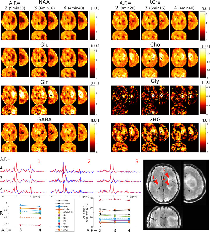
FIG. 4:
ECCENTRIC metabolic imaging in a WHO-3 Astrocytoma patient with IDH1(R132H) mutation (Patient #2 in Table I). The performance of 3D-ECCENTRIC 1H-FID-MRSI, acquired at 3.4 mm isotropic resolution with acceleration factors (AF) of 2, and retrospectively accelerated to 3 and 4, was compared. Top, metabolic images of eight metabolites for all accelerations. Bottom, spectra from three brain locations indicated by red arrows on the FLAIR image, correlation coefficient (R) between and , metabolite quantification error estimate (CRLB), linewidth (FWHM), and signal-to-noise ratio (SNR) across AF.

