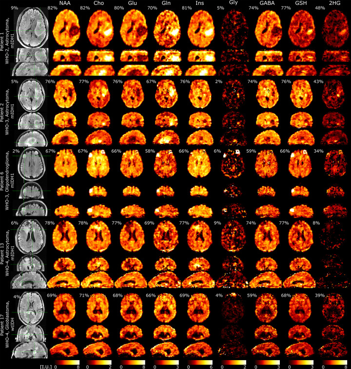
FIG. 5:
Metabolic imaging in glioma patients acquired with 3D-ECCENTRIC 1H-FID-MRSI at 3.4 mm isotropic resolution in 9min:20s accelerated by CS ). The top four patients have mutant IDH1(R132H) glioma: WHO-2 Astrocytoma, WHO-3 Astrocytoma, WHO-3 Oligodendroglioma, and WHO-4 Astrocytoma (Patients #1, #2, #6, #13 in Table I). The bottom fifth patient has a necrotic wild-type IDH1/2 WHO-4 Glioblastoma (Patient #17 in Table I). Intensity scale (I.U., institutional units) for each metabolite is the same across all patients. For each metabolite, we indicate the percentage of voxels inside the brain and FoV that meet the criteria of acceptable quality (CRLB < 20%, FWHM < 0.07ppm, SNR > 5). Percentage on FLAIR indicate the ratio of tumor volume to the total brain volume.

