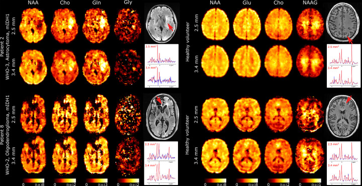
FIG. 7:
Ultra-high resolution metabolic imaging acquired with 3D-ECCENTRIC 1H-FID-MRSI at 2.5mm isotropic voxel size (, TA=10min : 26s) in two healthy volunteers and two glioma patients (Patients #2 and #13 in Table I). The ultra-high resolution metabolic imaging is compared to 3D-ECCENTRIC 1H-FID-MRSI at the typical voxel size of 3.4mm isotropic (, TA=9min : 20s). Two spectra from both spatial resolution and corresponding to the red arrow location are shown. The blue line represents the MRSI data and the red line is the fit performed by LCModel.

