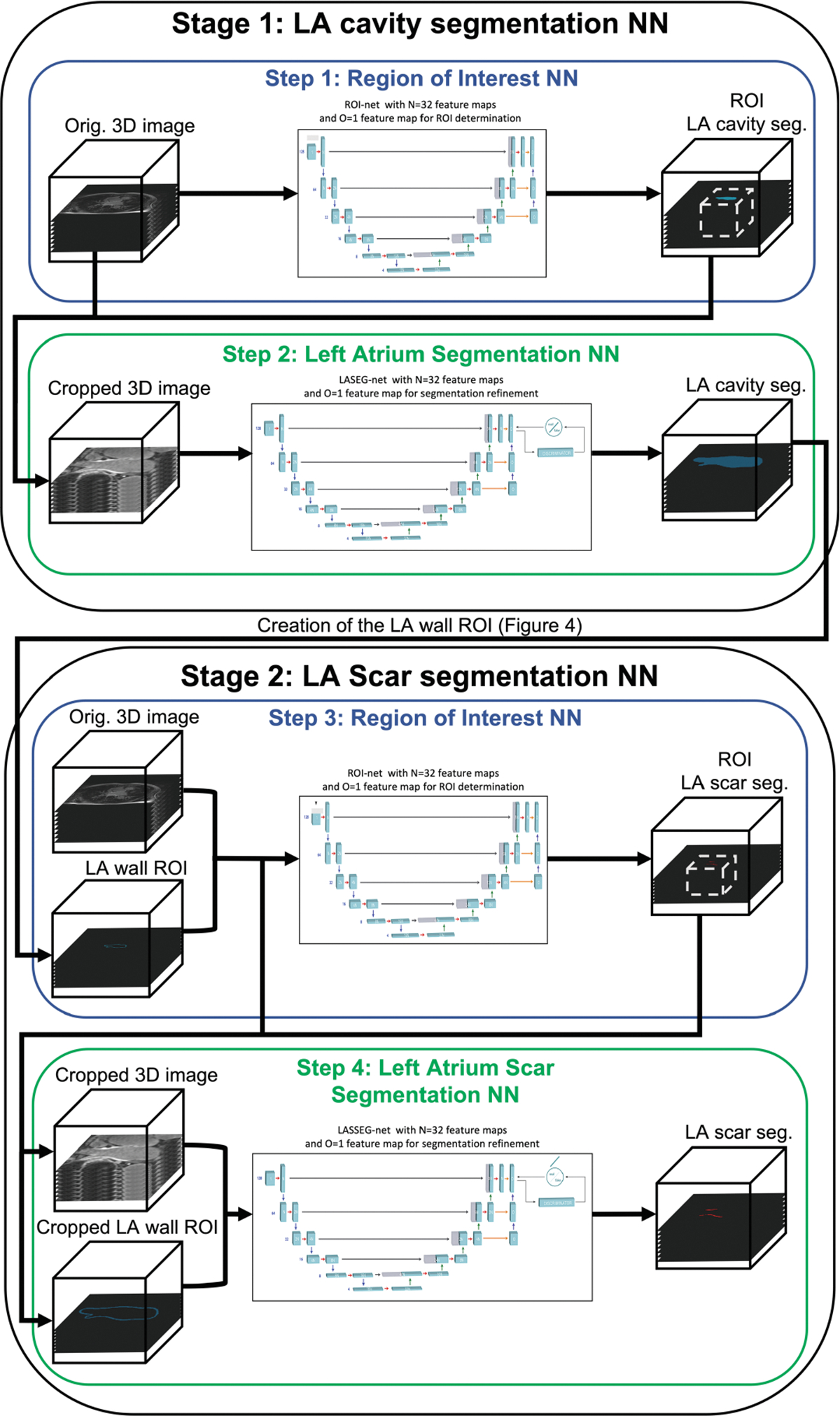
Fig. 3. LASSNet architecture.

Step 1: LGE-MRI images are passed into ROI NN which identifies the region where the left atrium is located. The image volume is then cropped to the ROI and resampled. Step 2: The image is inputted into the Left Atrium Segmentation Network (LASEG NN) to perform the LA cavity segmentation. LASEG NN includes a discriminator for adversarial training. Step 3: LGE-MRI images and LA wall ROI masks are passed into ROI NN which identifies the region where the left atrium scar is located. The image volume and the LA wall ROI are then cropped to the identified ROI and resampled. Step 4: Cropped LGE-MRI images and LA wall ROI masks are then inputted into the Left Atrium Scar Segmentation Network (LASSEG NN) to refine the left atrium scar segmentation. LASSEG NN includes a discriminator for adversarial training.
