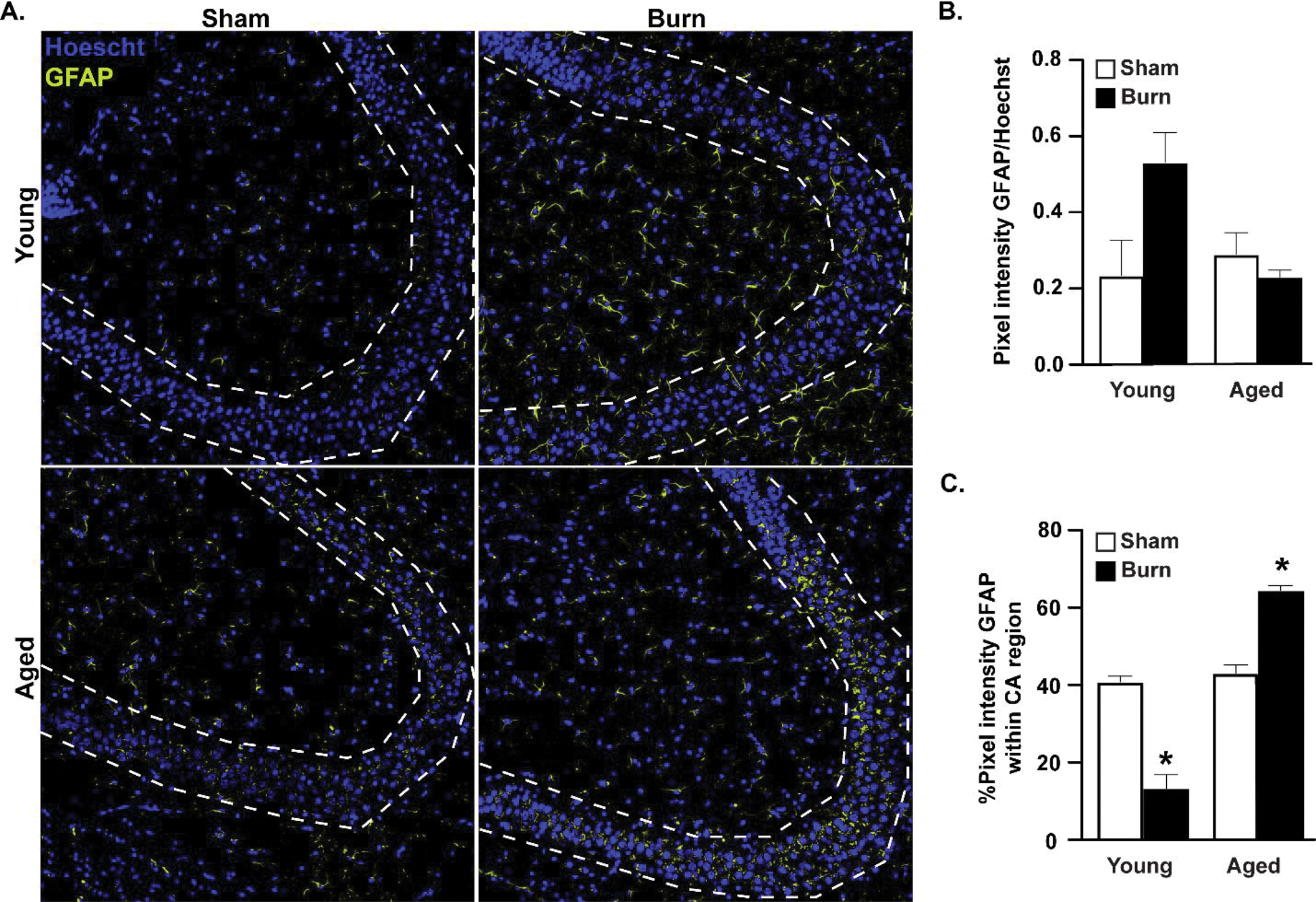
Figure 3. Increased GFAP+ reactive astrocytes in the CA3 region of the hippocampus of aged burn-injured mice.

(A) GFAP+ reactive astrocytes identified by immunofluorescence in perfused brains after burn injury. In aged burn-injured mice, GFAP+ cells increase their localization to the CA3 region (outlined in dotted line). Representative IF shown, n=3–4 mice per group, images shown from a representative of two independent experiments. (B) GFAP+ cells analyzed with ImageJ and presented as mean pixel density±SEM, normalized to Hoechst. n=3(sham)-4(burn) mice per group, experiment shown is a representative of two independent experiments. (C) Percentage of total GFAP+ signal that is within the CA3 (dotted lines) region reported as mean percentage±SEM. *p<0.05 from all other groups by ANOVA, Tukey’s post-hoc test. Graphs shown are from a single representative of two independent experiments. n=3 (sham) and 4 (burn) mice per group in this representative experiment.
