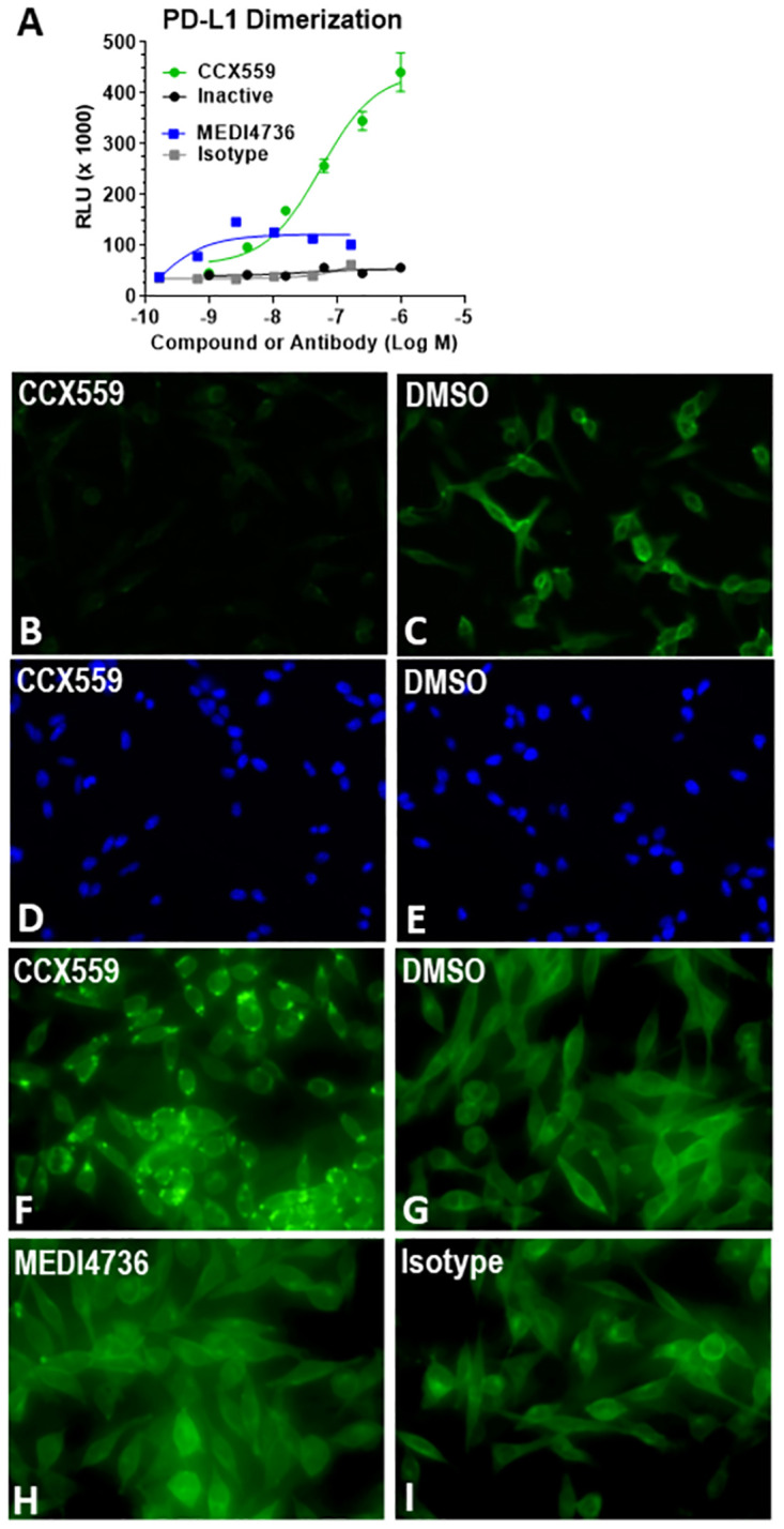
Fig 2. CCX559 induced human PD-L1 dimerization and relocalization to intracellular vesicles in vitro.

(A) In the PathHunter® PD-L1 Dimerization Assay, CCX559 (green circles) induced a dose-dependent dimerization signal, measured as RLU, but the inactive compound (black circles), MEDI4736 (blue squares) and the isotype control antibody (grey squares) had no or minimal effect. The error bars represent ± SD (n = 3). (B and C) Treatment of MC38-hPD-L1 cells for 5 hours with 1 μM CCX559 (B) reduced surface PD-L1 levels compared to a 0.1% DMSO vehicle control using the same exposure time (C). (D and E) The corresponding DAPI staining shows similar cell densities. (F through H) To detect intracellular PD-L1, MC38-hPD-L1 cells were permeabilized after treating with 300 nM CCX559 (F), 0.1% DMSO (G), 67 nM MEDI4736 (H) or isotype matched control antibody (I) for 22 hours. PD-L1 was observed in intracellular vesicles only in CCX559-treated cells. The images are representative of the entire cell field for each treatment and had identical exposure times.
