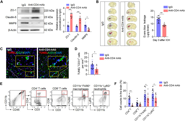
Fig. 8. Depletion of CD4+ T cells reduces BBB permeability and leucocyte infiltration in ICH mice.
C57BL/6 mice received intraperitoneal injection of 50 μg of anti-CD4 mAb or lgG control 24 hours before ICH induction with injection of autologous blood. (A) Brain tissue was collected at day 3 after ICH. Western blot shows tight junction protein level (claudin-5 and ZO-1) and MMP9 expression in ICH mice receiving anti-CD4 mAb or lgG control. Right: Quantification of ZO-1, claudin-5, and MMP9 in brain homogenates of ICH mice. n = 6 mice per group. Mann-Whitney test. (B) Concentrations of evans blue were measured at day 3 after ICH, with anti-CD4 mAb or IgG injection. n = 14 per group. Two-tailed unpaired Student’s t test. (C and D) Immunostaining (C) and quantification (D) of TUNEL in CD31+ endothelial cells of ICH mice received IgG control or anti-CD4 mAb separately. White dashed lines outline the hematoma area. Scale bars, 40 μm and (inset) 20 μm. n = 12 mice per group. Two-tailed unpaired Student’s t test. (E and F) Single-cell suspensions from brain tissue were collected at day 3 after ICH. (E) Gating strategy of brain-infiltrating neutrophils (CD11b+CD45hiLy6G+), monocytes/macrophages (CD11b+CD45hiF4/80+), CD4+ T cells (CD45hiCD3+CD4+), and CD8+ T cells (CD45hiCD3+CD8+) was shown. (F) Counts of the immune cell populations in ICH brains from mice receiving anti-CD4 mAb or IgG. n = 12 mice per group. Two-tailed unpaired Student’s t test. Means ± SD. *P < 0.05 and **P < 0.01.

