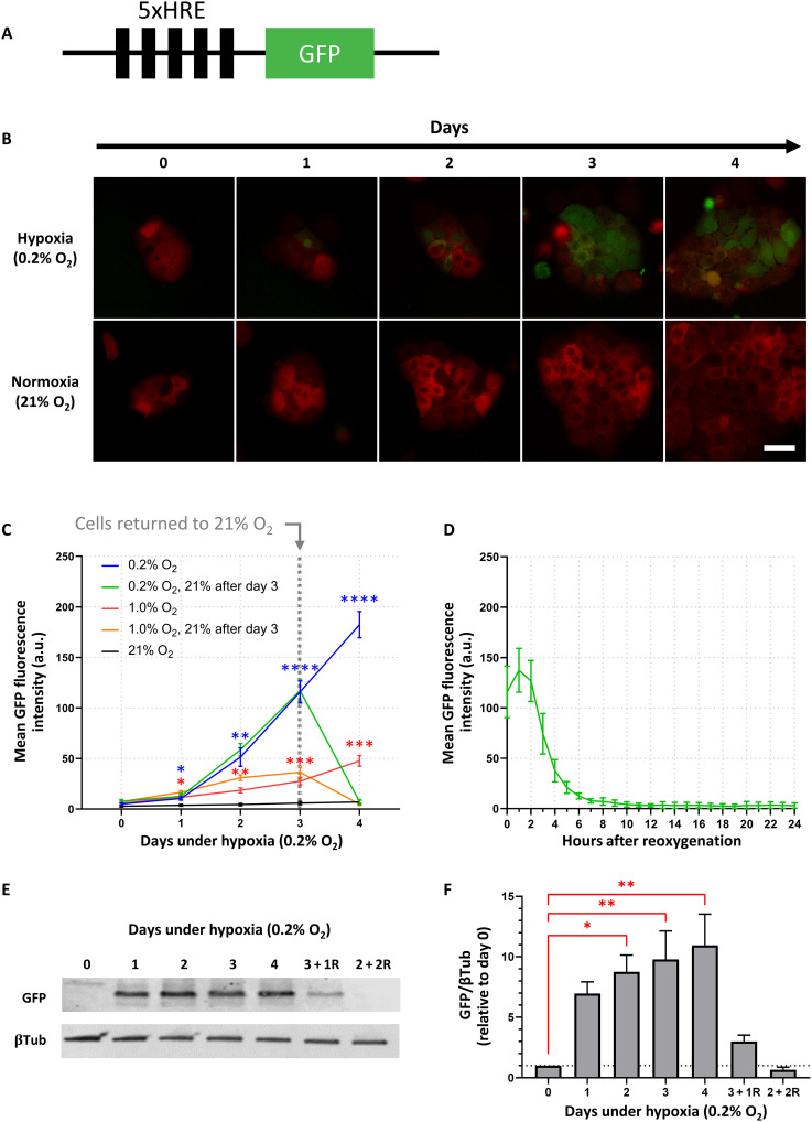
Fig. 1. Hypoxia drives 5xHRE-induced GFP expression in BxPC3-DsRed-5xHRE/GFP cells.
(A) Schematic of 5xHRE/GFP construct. (B) Representative images of BxPC3-DsRed-5xHRE/GFP cells growing under hypoxia (0.2% O2) and normoxia (21% O2) using in vitro live-cell fluorescence microscopy. Scale bar, 50 μm. (C) Quantification of GFP fluorescence intensity at 0.2, 1, and 21% O2. The green and orange lines indicate cells returned to 21% O2 between days 3 and 4. n = 9 replicates per group. (D) Hourly time lapse of the mean GFP fluorescence intensity after BxPC3-DsRed-5xHRE/GFP cells (previously incubated under 0.2% O2 for 3 days) were transferred to 21% O2 confirms the oxygen dependence of 5xHRE/GFP construct. n = 9 replicates per group. a.u., arbitrary units. (E) Western blot and (F) densitometric analysis of GFP expression [relative to β-tubulin (βTub)] after various time durations under 0.2% O2. Columns labeled with letter “R” indicate the number of days cells were reoxygenated at 21% O2 before protein collection. Data were obtained from three independent experiments. *P < 0.05, **P < 0.01, ***P < 0.001, and ****P < 0.0001.

