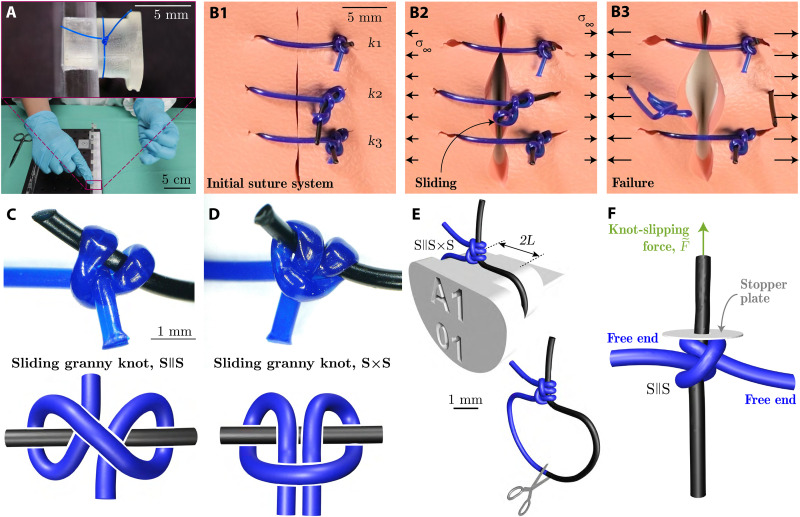
Fig. 1. Failure of surgical sliding knots.
(A) Photograph of the tying of a common sliding knot by an experienced surgeon in a Prolene polypropylene filament on a rigid support. (B1 to B3) Photographs illustrating knot safety and sliding for different levels of tightness of the S || S × S knot in a suture system on a practice pad, at increasing levels of the far-field stress, σ∞. (C and D) Optical microscope image (top) and topological diagram (bottom) of the S || S (C), and S × S (D) sliding-knot topologies. (E) Schematic of the S || S × S knot tied around a 3D-printed pin and visualization of the cutting location in the suture loop. (F) FEM-computed configuration for a S || S knot tied with a pre-tension of . The same configuration is implemented in the mechanical testing experiments to measure the slipping force, , of the S || S knot (cf. Fig. 2).

