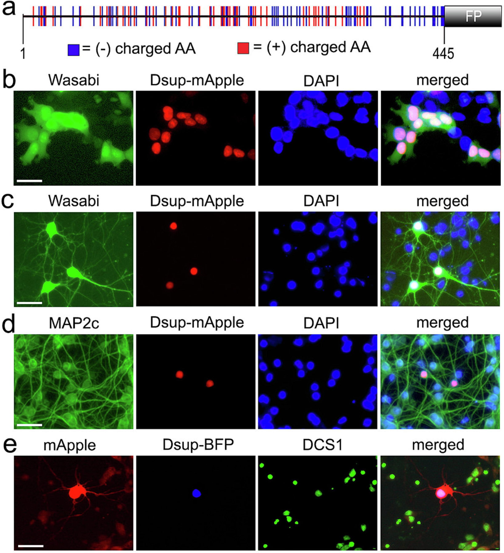
Figure 1.

Expression Dsup in primary neurons. (a) A scheme of the tardigrade Ramazzottius varieornatus Dsup protein, optimized for the expression in mammalian cells, fused to a fluorescent protein (mApple or BFP). The positions of acidic and basic amino acid residues within the 445 amino acid protein indicate that the Dsup protein is highly charged. (b) HEK 293 cells were co-transfected with Dsup-mApple and Wasabi. Cells were fixed 24 hours after transfection, stained the DAPI dye, and imaged. Note that the Dsup construct is nuclear. Scale bar, 10 µm. (c) Primary cortical neurons were co-transfected with Dsup-mApple and Wasabi. 24 hours after transfection, live cells were stained the DAPI dye and imaged. Note that Dsup-mApple is localized to the nucleus. Scale bar, 20 µm. (d) Primary cortical neurons transfected with Dsup-mApple, fixed 24 hours after transfection, and stained with the antibody against MAP2c and with the DAPI dye. Note that the Dsup-mApple construct is localized to the neuronal nucleus. Scale bar, 20 µm. (e) Primary cortical neurons were co-transfected with Dsup-BFP and mApple. 24 hours after transfection, neurons were fixed, stained the DCS1 dye and imaged. Note that Dsup-BFP is localized to the neuronal nucleus. Scale bar, 20 µm.
