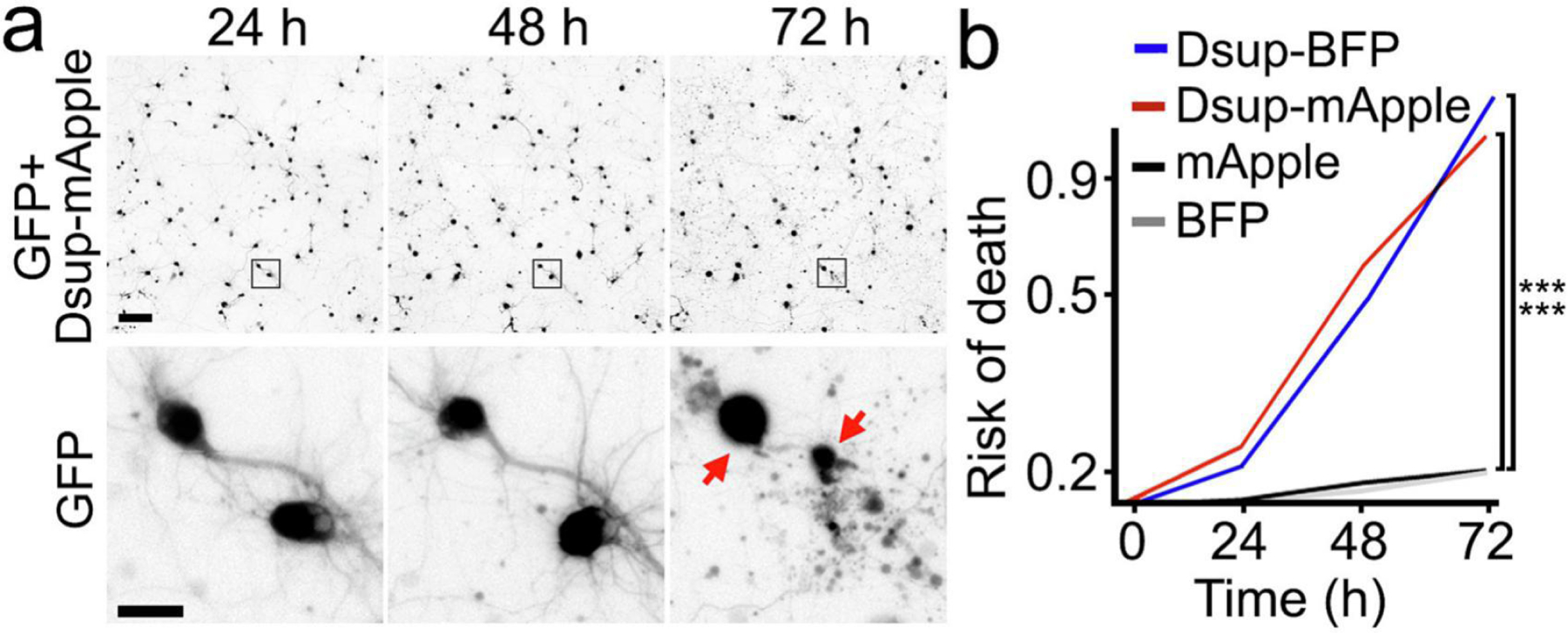
Figure 2.

The Dsup constructs are neurotoxic. (a) An example of longitudinal imaging of primary neurons. Cultured cortical neurons were co-transfected with Dsup-mApple and a green fluorescent protein (GFP) to visualize neuronal morphology. 24 hours after transfection, the same group of neurons were imaged longitudinally with an automated microscope at different time points. The top panel is a montage of non-overlapping images captured in one well of a 24-well plate. Scale bar is 100 µm. The bottom panel is zoomed onto two neurons (the GFP channel) to demonstrate longitudinal single-cell tracking. Scale bar is 10 µm. Note that both neurons died before the last imaging event (dead cells are depicted with red arrows). (b) Neurons were transfected with mApple and GFP or Dsup-mApple and GFP or with BFP and GFP or with Dsup-BFP and GFP, and tracked with an automated microscope for 72 hours. Risk of death curves demonstrate that Dsup-mApple and Dsup-BFP are toxic for neurons. ***p (mApple vs Dsup-mApple) < 0.0001, ***p (BFP vs Dsup-mBFP) < 0.0001 (log-rank test, JMP statistical software). More than one hundred neurons per group were analyzed from three independent experiments.
