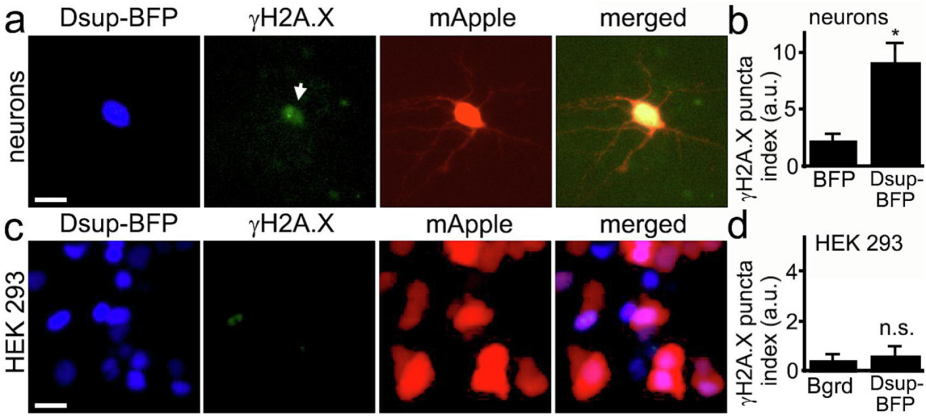
Figure 3. Dsup induces DNA DSBs in neurons.

(a) Primary neurons transfected with Dsup-BFP and mApple, fixed 48 hours after transfection, and immunostained with antibodies against γH2A.X. Note that the Dsup-BFP-positive neuron exhibits γH2AX puncta, indicating DNA DSBs (depicted with the arrow). Scale bar is 20 µm. (b) Images of fixed neurons from (a) were analyzed with ImageJ and the puncta index was estimated by measuring the standard deviation of γH2A.X fluorescence intensity. The puncta index of γH2A.X staining is increased in neurons that express Dsup-BFP. BFP, BFP-transfected neurons from Supplemental Figure 3. p= 0.0137 (t-test), a.u., arbitrary units. 271 Dsup-BFP-expressing and 321 BFP-expressing neurons were analyzed. Results were pooled from four experiments. (c) HEK 293 cells transfected with mApple and Dsup-BFP, fixed 48 hours after transfection, and immunostained with antibodies against γH2A.X. Virtually no nuclear γH2A.X puncta were observed in Dsup-BFP expressing cells. Scale bar is 10 µm. (d) Images of fixed HEK 293 cells from (c) were analyzed with ImageJ and the puncta index was estimated by measuring the standard deviation of γH2A.X fluorescence intensity and compared to background. Bgrd., background; n.s., non-significant, p=0.35 (t-test), a.u., arbitrary units. Results were pooled from three experiments.
