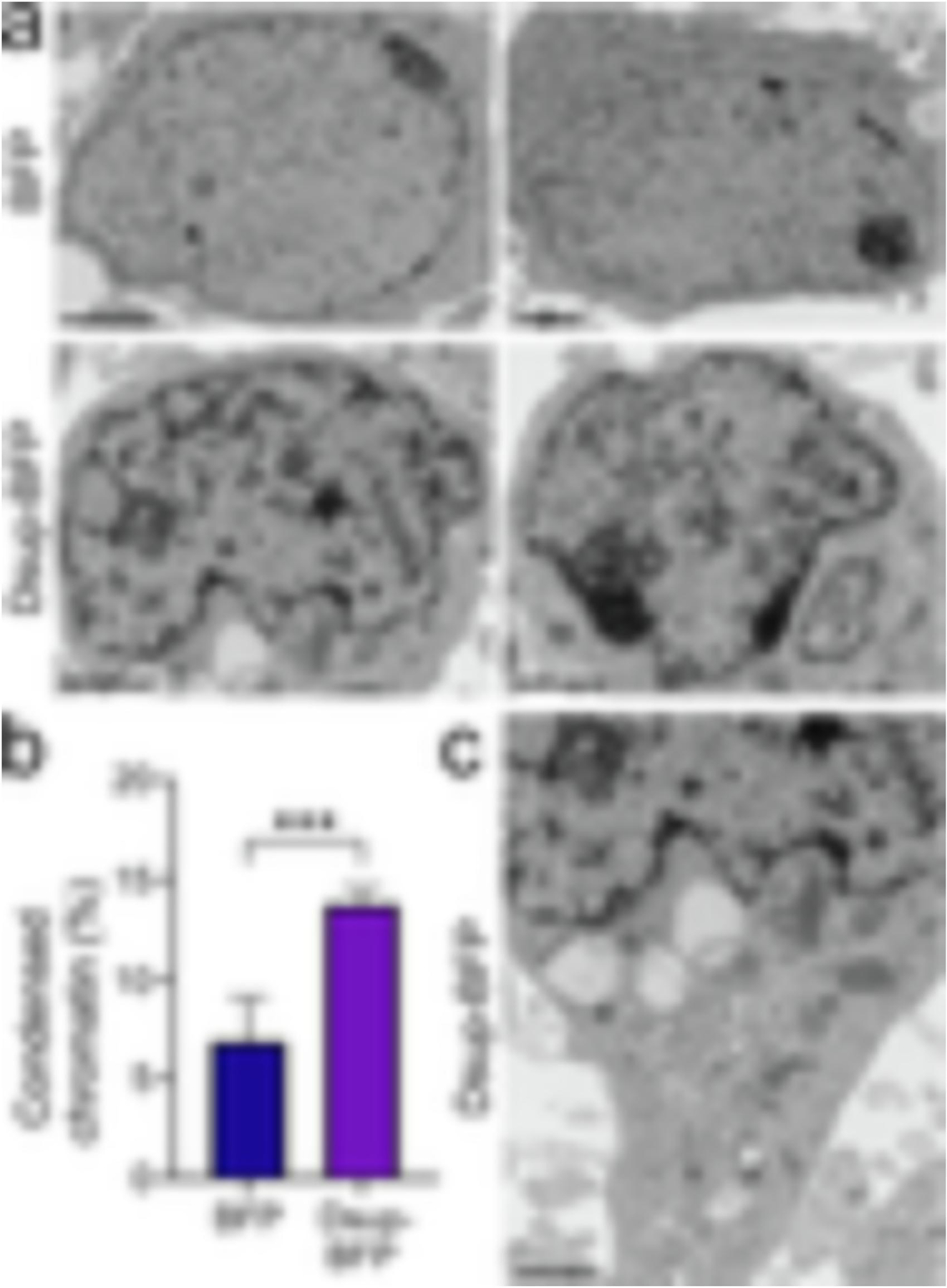
Figure 4. Dsup alters the structure of chromatin in primary neurons.

(a) Representative electron micrograph of cultured cortical neurons infected with the BFP lentivirus (BFP) or the Dsup-BFP lentivirus. Primary cultures were infected, fixed 72 hours when the expression plateaued and >70% of cells were expressing the constructs, and processed for electron microscopy imaging. Results were pooled from two experiments. Bar (BFP), 2 μm; bar (Dsup-BFP), 1 μm. (b) The percentage of condensed chromatin was calculated in the BFP-infected and Dsup-BFP-infected cohorts. P=0.0002 (t-test). (c) The cytoplasm of the neuron from the Dsup-BFP-infected cohort from (a) does not exhibit major abnormalities. Bar, 1 μm.
