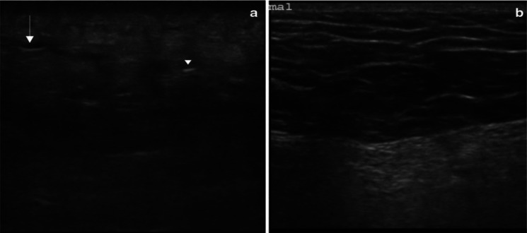
Fig. 5.
Ultrasonography of left gluteal soft tissue with a necrotizing fasciitis and b normal soft tissue on the contralateral side using a linear probe. In the affected tissue, the normal subcutaneous architecture is lost, and diffusely increased echogenicity is present. These hypoechoic regions correspond to little fluid accumulations (arrow) and a hyperechoic focus with posterior dirty acoustic shadowing, corresponding to gas in the soft tissue (arrowhead)—source: Magalhaes et al. with permission

