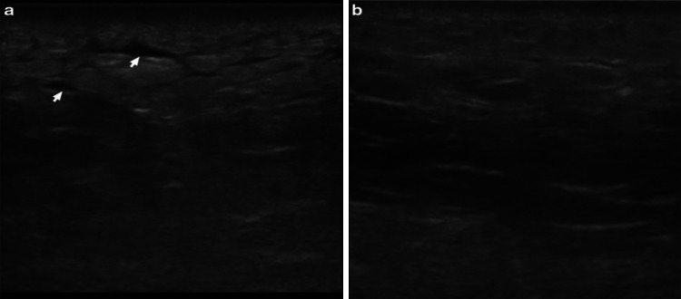
Fig. 6.
Ultrasonography of left gluteal soft tissue with a necrotizing fasciitis four days post-hospitalization and b 8 days post-hospitalization using a linear probe. These images show the progression of the sonographic appearance of necrotizing fasciitis, initially with an accumulation of fluid in the subcutaneous tissue (arrows), giving it a “cobblestone” appearance and a progressive return to the normal architecture of the subcutaneous tissue over time.
Source: Magalhaes et al. with permission

