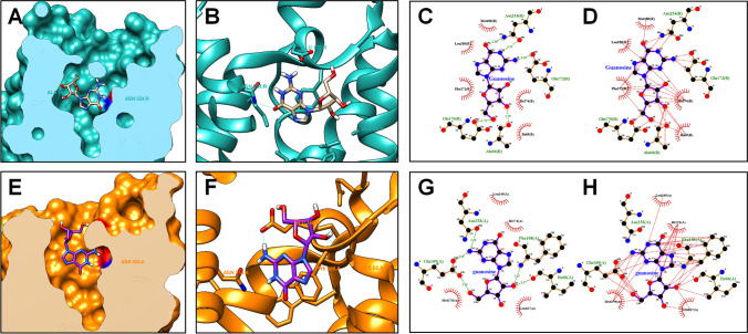
Fig. 7.
Molecular interactions between guanosine and A1R or A2AR. The putative binding site of guanosine to A1R (A). Structure of the A1R complexed with guanosine (B). Representative pose of guanosine complexed with A1R, in which guanosine–A2AR hydrogen bond are represented as green dashed lines (C). Representative pose of guanosine complexed with A1R, in which guanosine–A1R hydrophobic contacts are represented as red dashed lines (D). The putative binding site of guanosine to A2AR (E). Structure of the A2AR complexed with guanosine (F). Representative pose of guanosine complexed with A2AR, in which guanosine–A2AR hydrogen bonds are represented as green dashed lines (G). Representative pose of guanosine complexed with A2AR, in which guanosine–A1R hydrophobic contacts are represented as red dashed lines (H)

