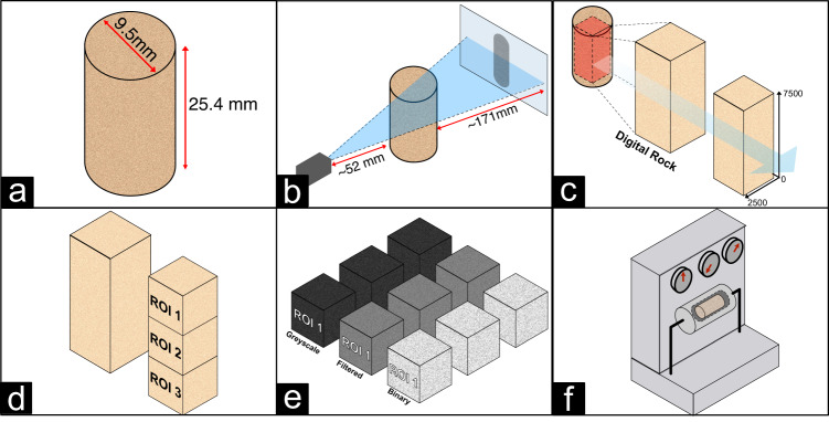
Fig. 1.
Conceptual overview of the rock sample study. (a) Schematic of a cylindrical rock plug sample having a length of 25.4 mm and a diameter of 9.5 mm. (b) Schematic of the X-Ray μCT imaging process. (c) Visualization of the image cube cropping process. (d) Data cube subdivision by regions of interest (ROI). (e) Data cube processing from greyscale to binary images. (f) Schematic representation of porosity and permeability measurements in the lab.

