
FIGURE 1.
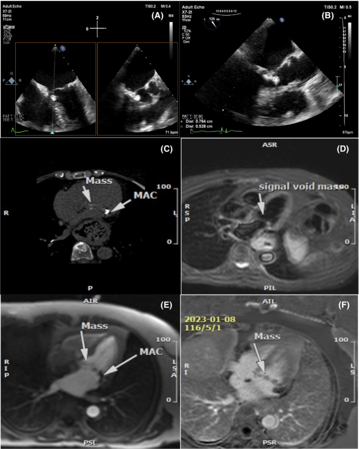
Multimodality imaging using transesophageal echocardiography (TEE), cardiac computed tomography (CT), and cardiac magnetic resonance imaging (CMR). TEE demonstrating a pedunculated mobile calcified mass (0.7 × 0.5 cm) attached to posterior of mitral valve (MV) annulus (A and B). Cardiac CT demonstrating mitral annular calcification (MAC) and calcified mass attached to MV annulus (C). CMR demonstrating the mass with low signal intensity on T2‐weighted images (D), with no perfusion in the first pass perfusion sequences (E) and no enhancement in the late‐enhancement sequences (F).