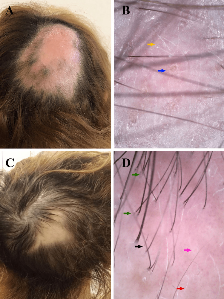
Figure 1. Clinical pictures and trichoscopy images before and after therapy.
Figure 1. A) Clinical picture before therapy: 15/8 cm sized bald patch in the right parietal area. B) Trichoscopy findings before therapy: yellow dots (blue arrow), short vellus hairs (yellow arrow). C) Clinical picture a month after the last treatment: 5/4 cm sized bald patch in the right parietal area. D) Trichoscopy findings a month after the last treatment: upright regrowing hairs (green arrows), vellus hairs (red arrow), minimal broken hairs (black arrow), and solitary yellow dots (pink arrow).

