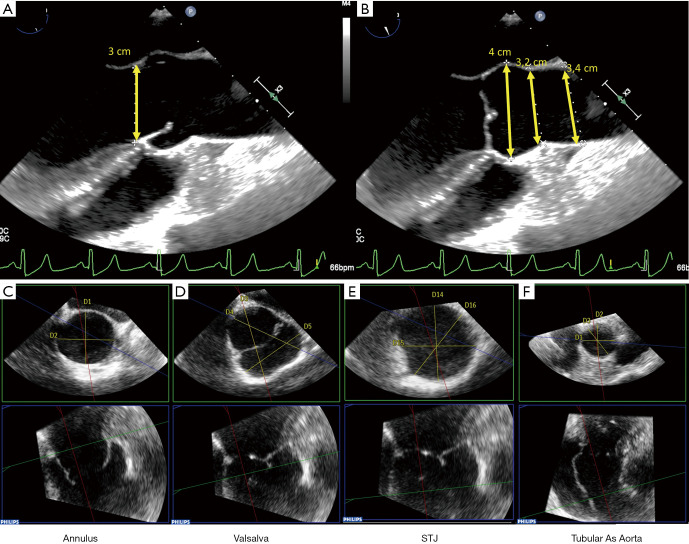
Figure 3.
The root phenotype is evaluated on a mid-esophageal 2D long-axis view. According to the guidelines of the American Society of Echocardiography and the European Association of Cardiovascular Imaging (26) the annulus is measured in systole (A) while the sinuses of Valsalva, the STJ and the ascending aorta are measured in diastole (B). The spatial resolution of modern 3D TEE allows the sonographer to use the inner edge-to-inner edge method. MPR provides additional information: the circular or elliptic shape of the annulus can be identified (C), a sinus asymmetry can be detected, and the largest diameter of the Valsalva can be measured (D), the precise location of the STJ, which is the upper border of the FAA, can be measured in different planes (E), and the tubular portion of the ascending aorta can be measured in different planes as well (F). STJ, sino-tubular junction; TEE, transesophageal echocardiography; MPR, multiplanar reconstruction; FAA, functional aortic annulus.

