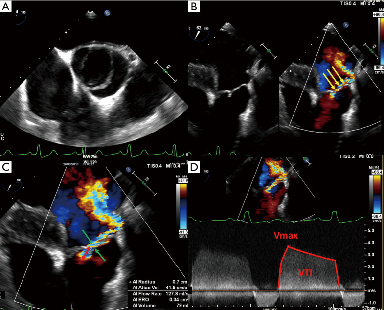
Figure 4.
Example of AR quantification using the PISA method in a patient with a BAV (fused left coronary cusp and right coronary cusp) (A). Deep transgastric long-axis transesophageal echocardiography color flow view of the eccentric AR (yellow arrows) (B). To calculate the flow rate across the regurgitant orifice, the aliasing velocity was decreased to 41.5 cm/s (velocity at the blue-red border), which increased the radius of flow convergence (r: red arrow 0.7 cm in this case). The vena contracta width can also be appreciated in this view (green arrows, 0.6 cm in this case) (C). This is used to calculate the regurgitant flow (= 2 × π × r2 × Valiasing), the EROA (regurgitant flow/maximal velocity), in this case 0.34 cm2, and the regurgitant volume (EROA × VTI), in this case 79 mL. Note that it can be difficult to align the Doppler beam to evaluate the regurgitant flow in the presence of a leaflet prolapse (D). AR, aortic regurgitation; PISA, proximal isovelocity surface area; BAV, bicuspid aortic valve; EROA, effective regurgitant orifice area; r, radius; VTI, velocity/time interval.

