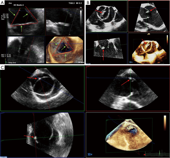
Figure 7.
Three-dimensional volume acquisition with multiplanar reconstruction allows for assessment of the quality of cusp tissue. (A) Example of systolic restrictive cusp motion: the tips of the cusps (green arrows) do not cross the virtual line (red dotted line) between two commissures in end-systole due to a root dilatation. (B) Example of thickening and calcification (red arrow) of the free margin, reducing the valve opening. (C) Example of a small calcification spot trapped into the non-fused cusp of a bicuspid aortic valve assessed by MPR (red arrow). MPR, multiplanar reconstruction.

