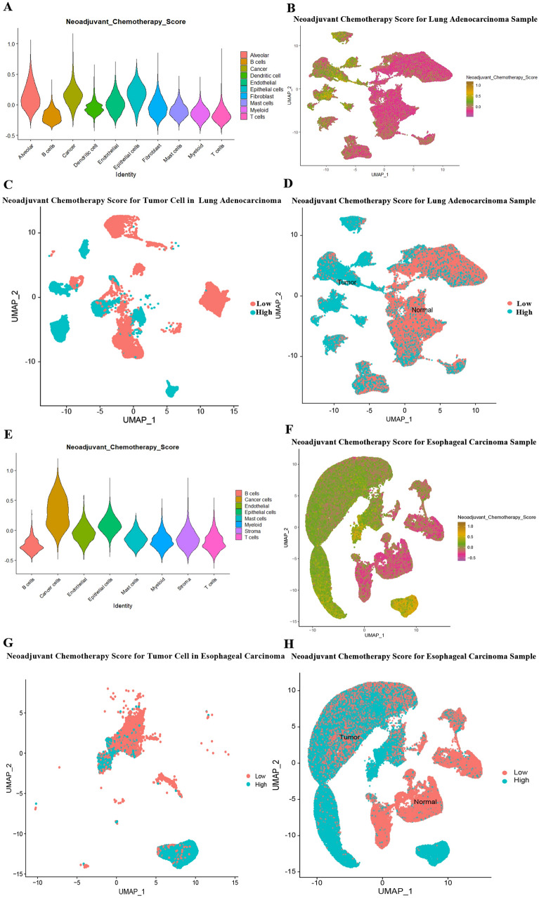
Fig. 4.
A, E: Violin plot showing the NCS values of each cell subpopulation in LUAD (A) and ESCC (E) tumors and normal tissues beside the tumors; B, F: UMAP plot showing the high and low NCS values of each cell in LUAD (B) and ESCC (F) tumors and normal tissues beside the tumors; C, G: Separating LUAD (C) and ESCC (G) tumor cells into two groups of high and low NCS; D, H: Tumor and peri-tumor normal tissues were separated according to UMAP, and all cells of LUAD (C) and ESCC (G) were divided into two groups according to the average value of NCS

