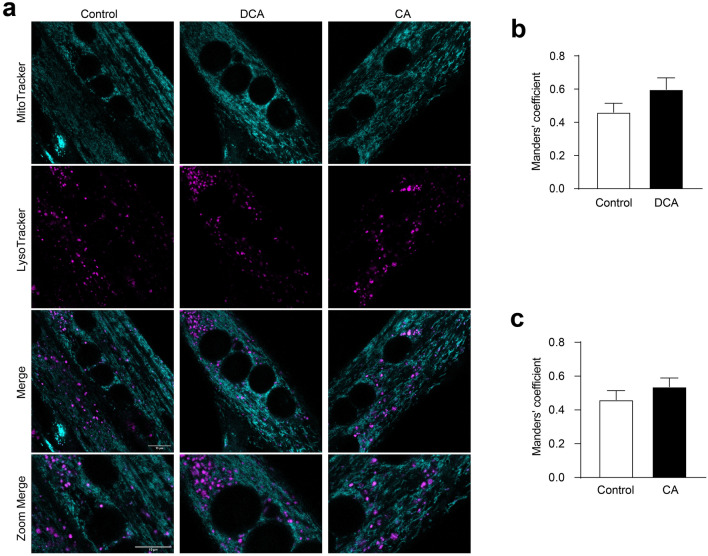
Fig. 4.
Mitophagy is unchanged by DCA and CA in C2C12 myotubes. a–c C2C12 myoblasts were differentiated for 5 days until forming myotubes. Then, myotubes were incubated with 120 μM of DCA or 500 μM of CA for 48 h. a After treatment with BA, cell culture was co-incubated with Mitotracker green FM (first panel) and Lysotracker Red DND-99 (second panel) probes. The co-localization (third panel and forth panel) between Mitotracker green and Lysotracker signals was determined as a parameter of mitophagy. Scale bar: 10 μm. Co-localization degree of fluorescent signals was calculated by Manders' coefficient using the JACoP plugin by ImageJ for (b) DCA and (c) CA. The values indicate the mean ± SEM of independent random fields. Scale bar:10 μm. (n = 3 independent experiments, *p < 0.05. t-test)

