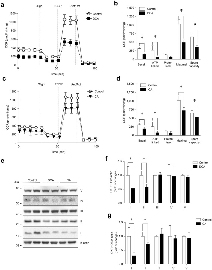
Fig. 6.
DCA and CA decrease the OCR and OXPHOS complexes I and II in C2C12 myotubes. Differentiated C2C12 cells forming myotubes were incubated with 120 μM of DCA (a–b) or 500 μM of CA (c-d) for 72 h. Basal, ATP-linked, H+-leak, maximal and spare OCR were determined as described in the Materials and Methods section. Values are expressed as pmol/min/mg and correspond to the mean ± SEM (n = 3, *p < 0.05. T-test). e Protein levels of I, II, III, and IV mitochondrial OXPHOS complexes were detected by Western blot analysis using β-actin levels as the loading control. Molecular weight markers are depicted in kDa. The quantitative value analysis is expressed as a fold of change for DCA (f) and CA (g). Values correspond to the mean ± SEM (n = 3 independent experiments, *p < 0.05. t-test)

